The MVK Monoclonal Antibody (CAB20906) is a high-quality antibody developed for reliable detection and analysis of target proteins. This antibody, raised in rabbits, offers high reactivity with human samples and has been validated for use in Western blot applications. By binding specifically to MVK, this antibody enables researchers to detect and analyze the protein in various cell types, making it ideal for studies in lipid metabolism and related disorders.MVK plays a crucial role in the mevalonate pathway, which is essential for the production of cholesterol, steroid hormones, and other important molecules in the body.
This antibody is validated for use in WB, ELISA applications and has demonstrated reactivity against Human samples.
Product Name:
MVK Monoclonal Antibody
SKU:
CAB20906
Size:
20μL, 100μL
Reactivity:
Human
Clone Number:
ARC2871
Conjugate:
Unconjugated
Immunogen:
Synthetic peptide. This information is considered to be commercially sensitive.
Recommended starting concentration is 1 μg/mL. Please optimize the concentration based on your specific assay requirements.
Synonyms:
MK, LRBP, MVLK, POROK3, MVK
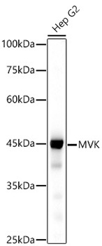
Positive Sample:
Hep G2
Cellular Localization:
Cytoplasm.
Calculated MW:
42kDa
Observed MW:
42kDa
This gene encodes the peroxisomal enzyme mevalonate kinase. Mevalonate is a key intermediate, and mevalonate kinase a key early enzyme, in isoprenoid and sterol synthesis. Mevalonate kinase deficiency caused by mutation of this gene results in mevalonic aciduria, a disease characterized psychomotor retardation, failure to thrive, hepatosplenomegaly, anemia and recurrent febrile crises. Defects in this gene also cause hyperimmunoglobulinaemia D and periodic fever syndrome, a disorder characterized by recurrent episodes of fever associated with lymphadenopathy, arthralgia, gastrointestinal dismay and skin rash. Alternative splicing results in multiple transcript variants.
Purification Method
Affinity purification
Gene ID
4598
Buffer Information
Store at -20℃. Avoid freeze / thaw cycles. Buffer: PBS containing 50% glycerol and 0.05% BSA, preserved with proclin300 or sodium azide, pH 7.3.
Western blot analysis of lysates from HepG2 cells, using MVK Rabbit mAb (CAB20906) at 1:500 dilution. Secondary antibody: HRP-conjugated Goat anti-Rabbit IgG (H+L) (CABS014) at 1:10000 dilution. Lysates/proteins: 25μg per lane. Blocking buffer: 3% nonfat dry milk in TBST. Detection: ECL Basic Kit (AbGn00020). Exposure time: 30s.
 at 1:10000 dilution. Lysates/proteins: 25ug per lane. Blocking buffer: 3% nonfat dry milk in TBST. Detection: ECL Basic Kit. Exposure time: 30s.")
 at 1:10000 dilution. Lysates/proteins: 25ug per lane. Blocking buffer: 3% nonfat dry milk in TBST. Detection: ECL Basic Kit. Exposure time: 30s.")

