The MyD88 Antibody (CAB0980) is a high-quality antibody developed for reliable detection and analysis of target proteins. This antibody, generated in rabbits, exhibits high specificity and sensitivity for detecting MYD88 in human samples, making it a reliable choice for Western blot applications. By binding to MYD88 protein, this antibody enables precise detection and analysis in various cell types, facilitating research in immunology and inflammation.MYD88 is a crucial player in the immune response, mediating signaling cascades that lead to the activation of inflammatory pathways.
This antibody is validated for use in WB, IF/ICC, ELISA applications and has demonstrated reactivity against Human, Mouse, Rat samples.
Product Name:
MyD88 Antibody
SKU:
CAB0980
Size:
20μL, 100μL
Reactivity:
Human, Mouse, Rat
Conjugate:
Unconjugated
Immunogen:
Synthetic peptide. This information is considered to be commercially sensitive.
Recommended starting concentration is 1 μg/mL. Please optimize the concentration based on your specific assay requirements.
Synonyms:
WM1, IMD68, MYD88D, MyD88
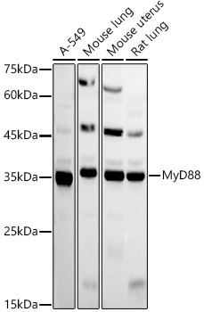
Positive Sample:
A-549, Mouse lung, Mouse uterus, Rat lung
Cellular Localization:
Cytoplasm.
Calculated MW:
33kDa
Observed MW:
33kDa
This gene encodes a cytosolic adapter protein that plays a central role in the innate and adaptive immune response. This protein functions as an essential signal transducer in the interleukin-1 and Toll-like receptor signaling pathways. These pathways regulate that activation of numerous proinflammatory genes. The encoded protein consists of an N-terminal death domain and a C-terminal Toll-interleukin1 receptor domain. Patients with defects in this gene have an increased susceptibility to pyogenic bacterial infections. Alternate splicing results in multiple transcript variants.
Purification Method
Affinity purification
Gene ID
4615
RRID
AB_2757499
Buffer Information
Store at -20℃. Avoid freeze / thaw cycles. Buffer: PBS with 0.09% Sodium azide,50% glycerol,pH7.3.
Western blot analysis of various lysates using MyD88 Rabbit pAb (CAB0980) at 1:500 dilution. Secondary antibody: HRP-conjugated Goat anti-Rabbit IgG (H+L) (CABS014) at 1:10000 dilution. Lysates/proteins: 25μg per lane. Blocking buffer: 3% nonfat dry milk in TBST. Detection: ECL Basic Kit (AbGn00020). Exposure time: 90s.
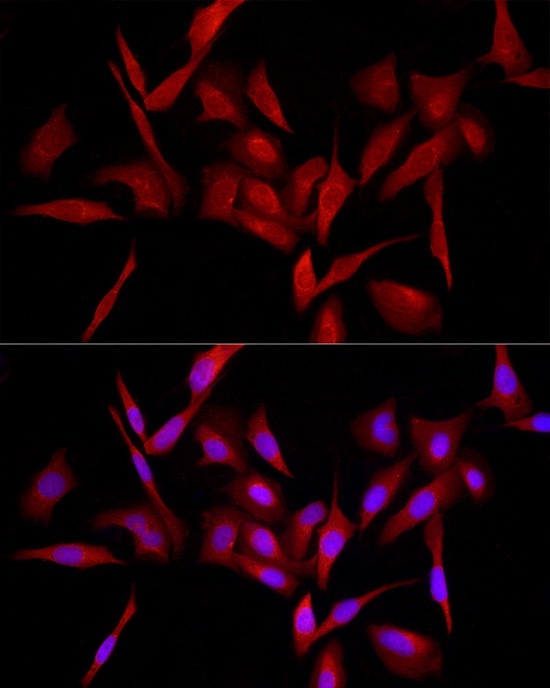
Immunofluorescence analysis of U2OS cells using MyD88 Rabbit pAb (CAB0980) at dilution of 1:100 (40x lens). Secondary antibody: Cy3-conjugated Goat anti-Rabbit IgG (H+L) (CABS007) at 1:500 dilution. Blue: DAPI for nuclear staining.


![[KO/KD Validated]MyD88 Rabbit Monoclonal Antibody (CAB22600)](https://cdn11.bigcommerce.com/s-h68l9z2lnx/images/stencil/590x590/products/223322/583859/kokd-validatedmyd88-rabbit-monoclonal-antibody__09830.1701185497.jpg?c=2 "[KO/KD Validated]MyD88 Rabbit Monoclonal Antibody (CAB22600)")