The MYEF2 Antibody (CAB15829) is a high-quality antibody developed for reliable detection and analysis of target proteins. This antibody, produced in rabbits, exhibits high reactivity with human samples and is validated for use in Western blot applications. By binding to the MYEF2 protein, this antibody enables accurate detection and analysis in a variety of cell types, making it an invaluable tool for studies in neuroscience and developmental biology.MYEF2 plays a crucial role in the regulation of gene expression during neuronal differentiation, making it a key player in the development and function of the central nervous system.
This antibody is validated for use in WB, IHC-P, IF/ICC, ELISA applications and has demonstrated reactivity against Human, Mouse, Rat samples.
Product Name:
MYEF2 Antibody
SKU:
CAB15829
Size:
20μL, 100μL
Reactivity:
Human, Mouse, Rat
Conjugate:
Unconjugated
Immunogen:
Recombinant protein (or fragment).This information is considered to be commercially sensitive.
Recommended starting concentration is 1 μg/mL. Please optimize the concentration based on your specific assay requirements.
Synonyms:
MEF-2, MST156, myEF-2, MSTP156, HsT18564, MYEF2
Positive Sample:
Jurkat, Mouse lung, Mouse testis, Mouse brain
Cellular Localization:
Nucleus.
Calculated MW:
64kDa
Observed MW:
64kDa
Enables RNA binding activity. Involved in myotube differentiation and neuron differentiation. Located in nucleus.
Purification Method
Affinity purification
Gene ID
50804
RRID
AB_2763253
Buffer Information
Store at -20℃. Avoid freeze / thaw cycles. Buffer: PBS with 0.01% thimerosal,50% glycerol,pH7.3.
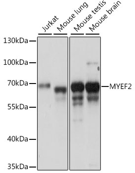
Western blot analysis of various lysates using MYEF2 Rabbit pAb (CAB15829) at 1:1000 dilution. Secondary antibody: HRP-conjugated Goat anti-Rabbit IgG (H+L) (CABS014) at 1:10000 dilution. Lysates/proteins: 25μg per lane. Blocking buffer: 3% nonfat dry milk in TBST. Detection: ECL Basic Kit (AbGn00020). Exposure time: 30s.
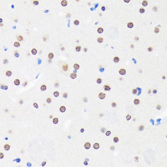
Immunohistochemistry analysis of paraffin-embedded Mouse brain using MYEF2 Rabbit pAb (CAB15829) at dilution of 1:100 (40x lens). Microwave antigen retrieval performed with 0.01M PBS Buffer (pH 7.2) prior to IHC staining.
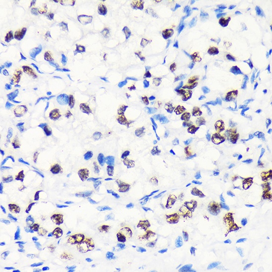
Immunohistochemistry analysis of paraffin-embedded Human liver cancer using MYEF2 Rabbit pAb (CAB15829) at dilution of 1:100 (40x lens). Microwave antigen retrieval performed with 0.01M PBS Buffer (pH 7.2) prior to IHC staining.
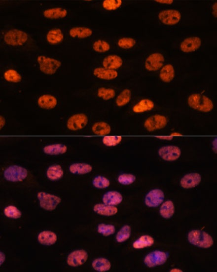
Immunofluorescence analysis of C6 cells using MYEF2 Rabbit pAb (CAB15829) at dilution of 1:100. Secondary antibody: Cy3-conjugated Goat anti-Rabbit IgG (H+L) (CABS007) at 1:500 dilution. Blue: DAPI for nuclear staining.