The MYH4 Antibody (CAB15293) is a high-quality antibody developed for reliable detection and analysis of target proteins. This antibody, produced in rabbits, is highly specific for MYH4 in human samples and is validated for use in Western blot applications. By binding to the MYH4 protein, this antibody allows for accurate detection and analysis in a variety of cell types, making it ideal for investigations in muscle biology and physiology.MYH4, a myosin heavy chain protein found in skeletal muscle, is essential for muscle contraction and movement.
This antibody is validated for use in WB, IHC-P, ELISA, IF-P applications and has demonstrated reactivity against Human, Mouse, Rat samples.
Product Name:
MYH4 Antibody
SKU:
CAB15293
Size:
20μL, 100μL
Reactivity:
Human, Mouse, Rat
Conjugate:
Unconjugated
Immunogen:
Synthetic peptide. This information is considered to be commercially sensitive.
Sequence:
ETLK RENK NLQQ EISD LTEQ IAEG GKHI HELE KVKK QLDH EKSE LQTS LEEA EASL EHEE GKIL RIQL ELNQ VKSE IDRK IAEK DEEL DQLK RNHL RVVE S
Tested Applications:
WBIHC-PELISAIF-P
Recommended Dilution:
WB
1:100 - 1:500
IF-P
1:50 - 1:200
IHC-P
1:50 - 1:200
ELISA
Recommended starting concentration is 1 μg/mL. Please optimize the concentration based on your specific assay requirements.
Synonyms:
MYH2B, MyHC-2B, MyHC-IIb, MYH4
Positive Sample:
Mouse skeletal muscle, Rat skeletal muscle
Cellular Localization:
Cytoplasm, Myofibril.
Calculated MW:
223kDa
Observed MW:
250kDa
Enables double-stranded RNA binding activity. Involved in muscle contraction. Located in myofibril.
Purification Method
Affinity purification
Gene ID
4622
RRID
AB_2762194
Buffer Information
Store at -20℃. Avoid freeze / thaw cycles. Buffer: PBS containing 50% glycerol, preserved with proclin300 or sodium azide, pH 7.3.
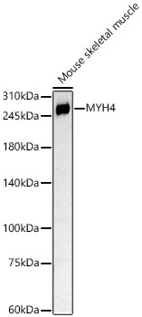
Western blot analysis of lysates from Mouse skeletal muscle, using MYH4 Rabbit pAb (CAB15293) at 1:400 dilution. Secondary antibody: HRP-conjugated Goat anti-Rabbit IgG (H+L) (CABS014) at 1:10000 dilution. Lysates/proteins: 25μg per lane. Blocking buffer: 3% nonfat dry milk in TBST. Detection: ECL Basic Kit (AbGn00020). Exposure time: 90s.
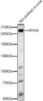
Western blot analysis of various lysates, using MYH4 Rabbit pAb (CAB15293) at 1:400 dilution. Secondary antibody: HRP-conjugated Goat anti-Rabbit IgG (H+L) (CABS014) at 1:10000 dilution. Lysates/proteins: 25μg per lane. Blocking buffer: 3% nonfat dry milk in TBST. Detection: ECL Enhanced Kit (AbGn00021). Exposure time: 60s.
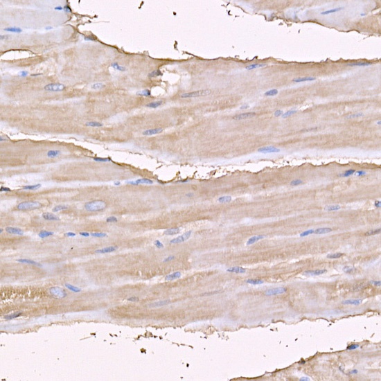
Immunohistochemistry analysis of paraffin-embedded Mouse skeletal muscle using MYH4 Rabbit pAb (CAB15293) at dilution of 1:100 (40x lens). High pressure antigen retrieval performed with 0.01M Citrate buffer (pH 6.0) prior to IHC staining.
Immunohistochemistry analysis of paraffin-embedded Rat heart using MYH4 Rabbit pAb (CAB15293) at dilution of 1:100 (40x lens). High pressure antigen retrieval performed with 0.01M Citrate buffer (pH 6.0) prior to IHC staining.
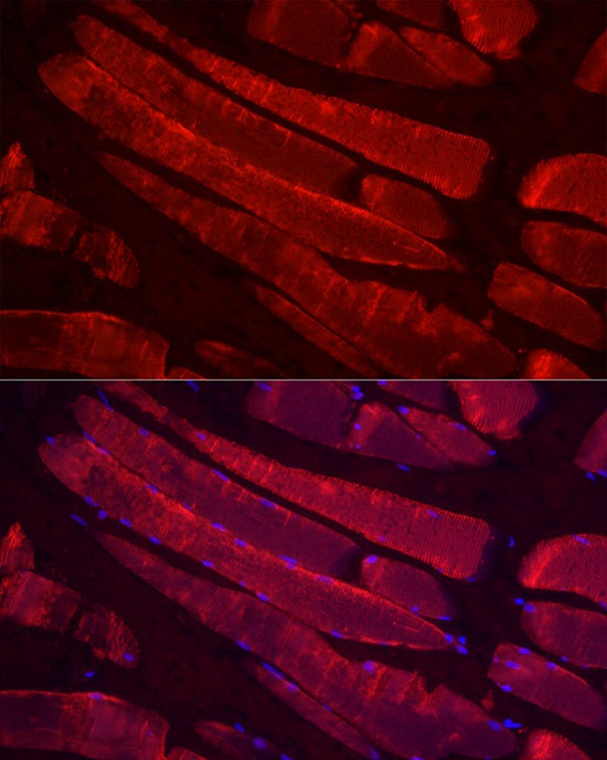
Immunofluorescence analysis of paraffin-embedded rat skeletal muscle using MYH4 Rabbit pAb (CAB15293) at dilution of 1:100 (40x lens). Secondary antibody: Cy3-conjugated Goat anti-Rabbit IgG (H+L) (CABS007) at 1:500 dilution. Blue: DAPI for nuclear staining.