The MYOM2 Antibody (CAB20526) is a high-quality antibody developed for reliable detection and analysis of target proteins. This antibody, produced in mice, is highly specific to Myom2 and is validated for use in immunofluorescence and immunohistochemistry applications.Myom2 is essential for maintaining the structure and function of muscle cells, playing a crucial role in muscle contraction and overall muscle physiology. Dysregulation of Myom2 has been linked to muscle disorders and myopathies, making it an important target for research in muscle biology and disease.
This antibody is validated for use in WB, ELISA applications and has demonstrated reactivity against Human, Mouse, Rat samples.
Product Name:
MYOM2 Antibody
SKU:
CAB20526
Size:
20μL, 100μL
Reactivity:
Human, Mouse, Rat
Conjugate:
Unconjugated
Immunogen:
Recombinant protein (or fragment).This information is considered to be commercially sensitive.
Recommended starting concentration is 1 μg/mL. Please optimize the concentration based on your specific assay requirements.
Synonyms:
TTNAP, MYOM2
Positive Sample:
Mouse heart, Mouse skeletal muscle, Rat heart, Rat skeletal muscle
Cellular Localization:
M Band, Mitochondrion.
Calculated MW:
165kDa
Observed MW:
165kDa
The giant protein titin, together with its associated proteins, interconnects the major structure of sarcomeres, the M bands and Z discs. The C-terminal end of the titin string extends into the M line, where it binds tightly to M-band constituents of apparent molecular masses of 190 kD and 165 kD. The predicted MYOM2 protein contains 1,465 amino acids. Like MYOM1, MYOM2 has a unique N-terminal domain followed by 12 repeat domains with strong homology to either fibronectin type III or immunoglobulin C2 domains. Protein sequence comparisons suggested that the MYOM2 protein and bovine M protein are identical.
Purification Method
Affinity purification
Gene ID
9172
Buffer Information
Store at -20℃. Avoid freeze / thaw cycles. Buffer: PBS with 0.01% thimerosal,50% glycerol,pH7.3.
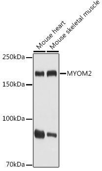
Western blot analysis of various lysates using MYOM2 Rabbit pAb (CAB20526) at 1:1000 dilution. Secondary antibody: HRP-conjugated Goat anti-Rabbit IgG (H+L) (CABS014) at 1:10000 dilution. Lysates/proteins: 25μg per lane. Blocking buffer: 3% nonfat dry milk in TBST. Detection: ECL Basic Kit (AbGn00020). Exposure time: 1s.
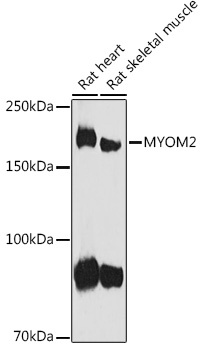
Western blot analysis of various lysates using MYOM2 Rabbit pAb (CAB20526) at 1:1000 dilution. Secondary antibody: HRP-conjugated Goat anti-Rabbit IgG (H+L) (CABS014) at 1:10000 dilution. Lysates/proteins: 25μg per lane. Blocking buffer: 3% nonfat dry milk in TBST. Detection: ECL Basic Kit (AbGn00020). Exposure time: 60s.