The PDE4DIP Antibody (CAB7765) is a high-quality antibody developed for reliable detection and analysis of target proteins. This antibody, produced in rabbits, is highly specific for human samples and is validated for use in Western blot applications. By binding to the Myomegalin protein, this antibody allows for detection and analysis in a variety of cell types, making it an ideal choice for studies in cell biology and cancer research.
This antibody is validated for use in WB, ELISA applications and has demonstrated reactivity against Mouse samples.
Product Name:
PDE4DIP Antibody
SKU:
CAB7765
Size:
20μL, 100μL
Reactivity:
Mouse
Conjugate:
Unconjugated
Immunogen:
Recombinant protein (or fragment).This information is considered to be commercially sensitive.
The protein encoded by this gene serves to anchor phosphodiesterase 4D to the Golgi/centrosome region of the cell. Defects in this gene may be a cause of myeloproliferative disorder (MBD) associated with eosinophilia. Several transcript variants encoding different isoforms have been found for this gene.
Purification Method
Affinity purification
Gene ID
9659
RRID
AB_2770827
Buffer Information
Store at -20℃. Avoid freeze / thaw cycles. Buffer: PBS containing 50% glycerol, preserved with proclin300 or sodium azide, pH 7.3.
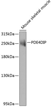
Western blot analysis of lysates from mouse skeletal muscle, using PDE4DIP Rabbit pAb (CAB7765) at 1:1000 dilution. Secondary antibody: HRP-conjugated Goat anti-Rabbit IgG (H+L) (CABS014) at 1:10000 dilution. Lysates/proteins: 25μg per lane. Blocking buffer: 3% nonfat dry milk in TBST. Detection: ECL Enhanced Kit (AbGn00021). Exposure time: 60s.