The MYH1 Antibody (CAB6935) is a high-quality antibody developed for reliable detection and analysis of target proteins. This antibody, produced in rabbits, exhibits high reactivity with human samples and is specifically validated for Western blot applications.Myosin-1 is a key player in cell movement and shape changes, making it essential for normal cellular function. By targeting the myosin-1 protein, this antibody enables precise detection and analysis of this important molecule in a variety of cell types.
This antibody is validated for use in WB, IHC-P, ELISA, IF-P applications and has demonstrated reactivity against Human, Mouse, Rat samples.
Product Name:
MYH1 Antibody
SKU:
CAB6935
Size:
20μL, 100μL
Reactivity:
Human, Mouse, Rat
Conjugate:
Unconjugated
Immunogen:
Recombinant protein (or fragment).This information is considered to be commercially sensitive.
Recommended starting concentration is 1 μg/mL. Please optimize the concentration based on your specific assay requirements.
Synonyms:
MYHa, HEL71, MYHSA1, MyHC-2x, MyHC-2X/D, MYH1
Positive Sample:
Mouse skeletal muscle, Rat skeletal muscle, Mouse skeletal muscle
Cellular Localization:
Cytoplasm, Myofibril.
Calculated MW:
223kDa
Observed MW:
251kDa/250kDa
Myosin is a major contractile protein which converts chemical energy into mechanical energy through the hydrolysis of ATP. Myosin is a hexameric protein composed of a pair of myosin heavy chains (MYH) and two pairs of nonidentical light chains. Myosin heavy chains are encoded by a multigene family. In mammals at least 10 different myosin heavy chain (MYH) isoforms have been described from striated, smooth, and nonmuscle cells. These isoforms show expression that is spatially and temporally regulated during development.
Purification Method
Affinity purification
Gene ID
4619
RRID
AB_2767493
Buffer Information
Store at -20℃. Avoid freeze / thaw cycles. Buffer: PBS containing 50% glycerol, preserved with proclin300 or sodium azide, pH 7.3.
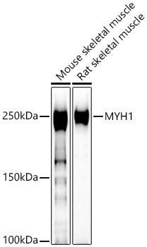
Western blot analysis of various lysates using MYH1 Rabbit pAb (CAB6935) at 1:400 dilution. Secondary antibody: HRP-conjugated Goat anti-Rabbit IgG (H+L) (CABS014) at 1:10000 dilution. Lysates/proteins: 25μg per lane. Blocking buffer: 3% nonfat dry milk in TBST. Detection: ECL Basic Kit (AbGn00020). Exposure time: 30s.
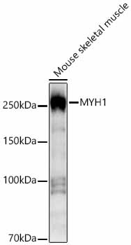
Western blot analysis of lysates from Mouse skeletal muscle using MYH1 Rabbit pAb (CAB6935) at 1:1000 dilution. Secondary antibody: HRP-conjugated Goat anti-Rabbit IgG (H+L) (CABS014) at 1:10000 dilution. Lysates/proteins: 25 μg per lane. Blocking buffer: 3% nonfat dry milk in TBST. Detection: ECL Basic Kit (AbGn00020). Exposure time:3s.
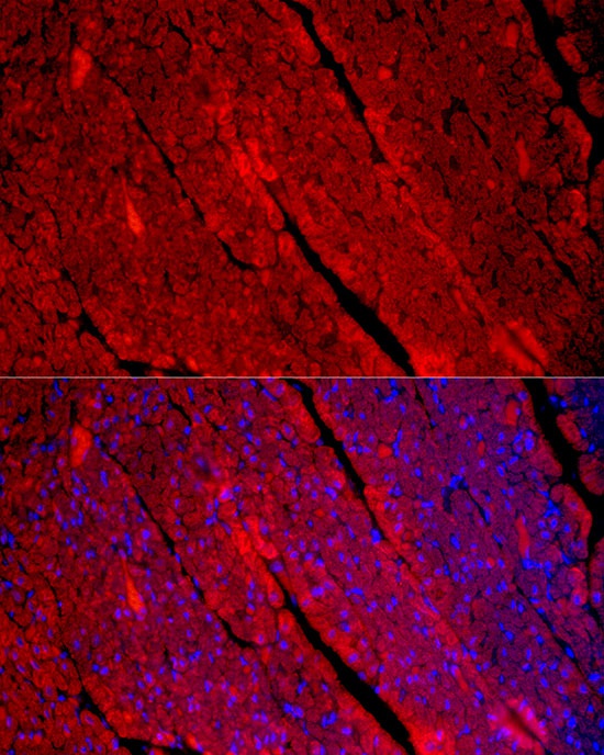
Immunofluorescence analysis of paraffin-embedded Human heart using MYH1 Rabbit pAb (CAB6935) at dilution of 1:100 (40x lens). Secondary antibody: Cy3-conjugated Goat anti-Rabbit IgG (H+L) (CABS007) at 1:500 dilution. Blue: DAPI for nuclear staining.
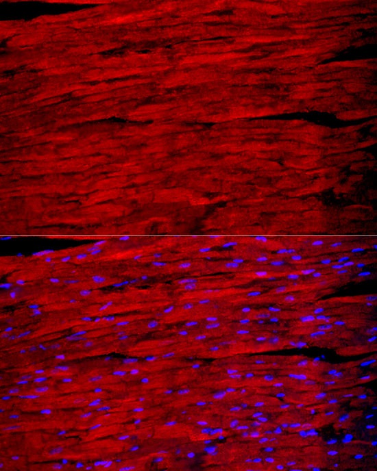
Immunofluorescence analysis of paraffin-embedded mouse heart using MYH1 Rabbit pAb (CAB6935) at dilution of 1:100 (40x lens). Secondary antibody: Cy3-conjugated Goat anti-Rabbit IgG (H+L) (CABS007) at 1:500 dilution. Blue: DAPI for nuclear staining.