The N4BP1 Antibody (CAB8474) is a high-quality antibody developed for reliable detection and analysis of target proteins. This antibody, generated in rabbits, exhibits high reactivity with human samples and has been validated for use in Western blot applications.N4BP1 is known to interact with multiple proteins involved in cell growth and proliferation, suggesting its potential role in cancer development and progression. By targeting N4BP1 with this antibody, researchers can study its function and mechanisms in different cell types, aiding in the advancement of cancer research and drug discovery efforts.
This antibody is validated for use in WB, IF/ICC, ELISA applications and has demonstrated reactivity against Human, Mouse samples.
Product Name:
N4BP1 Antibody
SKU:
CAB8474
Size:
20μL, 100μL
Reactivity:
Human, Mouse
Conjugate:
Unconjugated
Immunogen:
Recombinant protein (or fragment).This information is considered to be commercially sensitive.
Recommended starting concentration is 1 μg/mL. Please optimize the concentration based on your specific assay requirements.
Synonyms:
N4BP1
Positive Sample:
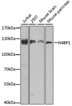
Jurkat, 293T, Mouse brain, Mouse pancreas
Cellular Localization:
Nucleus, Pml Body, Nucleolus.
Calculated MW:
100kDa
Observed MW:
120kDa
Predicted to enable mRNA binding activity; ribonuclease activity; and ubiquitin binding activity. Involved in cellular response to UV; negative regulation of macromolecule metabolic process; and regulation of innate immune response. Located in PML body; cytosol; and nucleolus. Orthologous to human N4BP1 (NEDD4 binding protein 1).
Purification Method
Affinity purification
Gene ID
80750
RRID
AB_2770510
Buffer Information
Store at -20℃. Avoid freeze / thaw cycles. Buffer: PBS, preserved with proclin300 or sodium azide, pH 7.3.
Western blot analysis of various lysates using N4BP1 Rabbit pAb (CAB8474) at 1:1000 dilution. Secondary antibody: HRP-conjugated Goat anti-Rabbit IgG (H+L) (CABS014) at 1:10000 dilution. Lysates/proteins: 25μg per lane. Blocking buffer: 3% nonfat dry milk in TBST. Detection: ECL Basic Kit (AbGn00020). Exposure time: 30s.
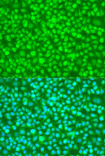
Immunofluorescence analysis of U2OS cells using N4BP1 Rabbit pAb (CAB8474) at dilution of 1:100. Secondary antibody: Cy3-conjugated Goat anti-Rabbit IgG (H+L) (CABS007) at 1:500 dilution. Blue: DAPI for nuclear staining.