The NAP1L5 Antibody (CAB17849) is a high-quality antibody developed for reliable detection and analysis of target proteins. This rabbit-derived antibody is highly specific to human samples and has been validated for use in Western blot applications. By binding to the NAP1L5 protein, this antibody allows for accurate detection and analysis in a variety of cell types.NAP1L5, also known as nucleosome assembly protein 1 like 5, plays a crucial role in regulating chromatin structure and facilitating the assembly of nucleosomes during DNA replication. Understanding the function of NAP1L5 is essential for unraveling the complexities of DNA packaging and gene expression.
This antibody is validated for use in WB, ELISA applications and has demonstrated reactivity against Human, Mouse samples.
Product Name:
NAP1L5 Antibody
SKU:
CAB17849
Size:
20μL, 100μL
Reactivity:
Human, Mouse
Conjugate:
Unconjugated
Immunogen:
Recombinant protein (or fragment).This information is considered to be commercially sensitive.
Recommended starting concentration is 1 μg/mL. Please optimize the concentration based on your specific assay requirements.
Synonyms:
DRLM, NAP1L5
Positive Sample:
SH-SY5Y, Mouse brain
Cellular Localization:
Nucleus.
Calculated MW:
20kDa
Observed MW:
20kDa/30kDa
This gene encodes a protein that shares sequence similarity to nucleosome assembly factors, but may be localized to the cytoplasm rather than the nucleus. Expression of this gene is downregulated in hepatocellular carcinomas. This gene is located within a differentially methylated region (DMR) and is imprinted and paternally expressed. There is a related pseudogene on chromosome 4.
Purification Method
Affinity purification
Gene ID
266812
RRID
AB_2770520
Buffer Information
Store at -20℃. Avoid freeze / thaw cycles. Buffer: PBS with 0.01% thimerosal,50% glycerol,pH7.3.
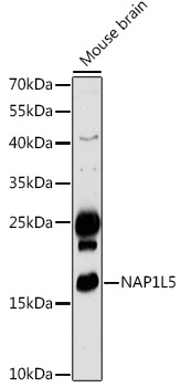
Western blot analysis of lysates from Mouse brain, using NAP1L5 Rabbit pAb (CAB17849) at 1:1000 dilution. Secondary antibody: HRP-conjugated Goat anti-Rabbit IgG (H+L) (CABS014) at 1:10000 dilution. Lysates/proteins: 25μg per lane. Blocking buffer: 3% nonfat dry milk in TBST. Detection: ECL Basic Kit (AbGn00020). Exposure time: 180s.
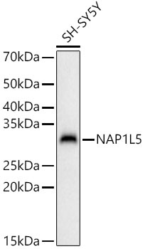
Western blot analysis of lysates from SH-SY5Y cells, using NAP1L5 Rabbit pAb (CAB17849) at 1:1000 dilution. Secondary antibody: HRP-conjugated Goat anti-Rabbit IgG (H+L) (CABS014) at 1:10000 dilution. Lysates/proteins: 25μg per lane. Blocking buffer: 3% nonfat dry milk in TBST. Detection: ECL Basic Kit (AbGn00020). Exposure time: 180s.