The NASP Antibody (CAB6938) is a high-quality antibody developed for reliable detection and analysis of target proteins. This antibody, raised in rabbits, is highly specific and reacts with NASP from various species, including human, mouse, and rat samples. Validated for use in immunofluorescence and immunoprecipitation assays, this antibody allows for the detection and analysis of NASP in a variety of cell types.NASP plays a crucial role in facilitating the assembly of nucleosomes, which are the building blocks of chromatin. By studying NASP, researchers can gain insights into the regulation of gene expression, DNA replication, and DNA repair processes.
This antibody is validated for use in WB, IHC-P, ELISA applications and has demonstrated reactivity against Human, Mouse, Rat samples.
Product Name:
NASP Antibody
SKU:
CAB6938
Size:
20μL, 100μL
Reactivity:
Human, Mouse, Rat
Conjugate:
Unconjugated
Immunogen:
Recombinant protein (or fragment).This information is considered to be commercially sensitive.
Recommended starting concentration is 1 μg/mL. Please optimize the concentration based on your specific assay requirements.
Synonyms:
HMDRA1, FLB7527, PRO1999, NASP
Positive Sample:
SW620, HL-60, 293T
Cellular Localization:
Cytoplasm, Nucleus.
Calculated MW:
85kDa
Observed MW:
85kDa
This gene encodes a H1 histone binding protein that is involved in transporting histones into the nucleus of dividing cells. Multiple isoforms are encoded by transcript variants of this gene. The somatic form is expressed in all mitotic cells, is localized to the nucleus, and is coupled to the cell cycle. The testicular form is expressed in embryonic tissues, tumor cells, and the testis. In male germ cells, this protein is localized to the cytoplasm of primary spermatocytes, the nucleus of spermatids, and the periacrosomal region of mature spermatozoa.
Purification Method
Affinity purification
Gene ID
4678
RRID
AB_2767496
Buffer Information
Store at -20℃. Avoid freeze / thaw cycles. Buffer: PBS containing 50% glycerol, preserved with proclin300 or sodium azide, pH 7.3.
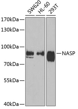
Western blot analysis of various lysates using NASP Rabbit pAb (CAB6938) at 1:1000 dilution. Secondary antibody: HRP-conjugated Goat anti-Rabbit IgG (H+L) (CABS014) at 1:10000 dilution. Lysates/proteins: 25μg per lane. Blocking buffer: 3% nonfat dry milk in TBST. Detection: ECL Basic Kit (AbGn00020). Exposure time: 90s.
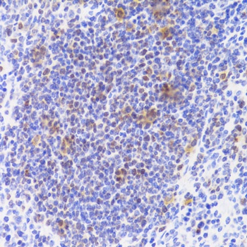
Immunohistochemistry analysis of paraffin-embedded Rat spleen using NASP Rabbit pAb (CAB6938) at dilution of 1:100 (40x lens). Microwave antigen retrieval performed with 0.01M PBS Buffer (pH 7.2) prior to IHC staining.
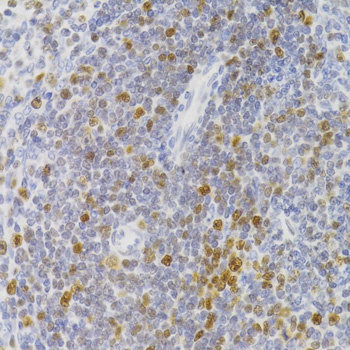
Immunohistochemistry analysis of paraffin-embedded Mouse spleen using NASP Rabbit pAb (CAB6938) at dilution of 1:100 (40x lens). Microwave antigen retrieval performed with 0.01M PBS Buffer (pH 7.2) prior to IHC staining.