The NAT8 Antibody (CAB7759) is a high-quality antibody developed for reliable detection and analysis of target proteins. This antibody, raised in rabbits, is highly reactive with human samples and has been validated for use in Western blot applications. NAT8, also known as N-acetyltransferase 8, has been implicated in conditions such as neurodegenerative diseases, kidney disorders, and metabolic syndromes. Research into the function and regulation of NAT8 is crucial for understanding its role in these conditions and developing potential therapies.
This antibody is validated for use in WB, IHC-P, ELISA applications and has demonstrated reactivity against Human, Mouse, Rat samples.
Product Name:
NAT8 Antibody
SKU:
CAB7759
Size:
20μL, 100μL
Reactivity:
Human, Mouse, Rat
Conjugate:
Unconjugated
Immunogen:
Recombinant protein (or fragment).This information is considered to be commercially sensitive.
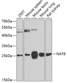
293T, Mouse spleen, Mouse testis, Mouse lung, Rat kidney
Cellular Localization:
Endoplasmic Reticulum Membrane, Endoplasmic Reticulum-Golgi Intermediate Compartment Membrane, Single-Pass Type Ii Membrane Protein.
Calculated MW:
26kDa
Observed MW:
26kDa
This gene, isolated using the differential display method to detect tissue-specific genes, is specifically expressed in kidney and liver. The encoded protein shows amino acid sequence similarity to N-acetyltransferases. A similar protein in Xenopus affects cell adhesion and gastrulation movements, and may be localized in the secretory pathway. A highly similar paralog is found in a cluster with this gene.
Purification Method
Affinity purification
Gene ID
9027
RRID
AB_2770523
Buffer Information
Store at -20℃. Avoid freeze / thaw cycles. Buffer: PBS containing 50% glycerol, preserved with proclin300 or sodium azide, pH 7.3.
Western blot analysis of various lysates using NAT8 Rabbit pAb (CAB7759) at 1:500 dilution. Secondary antibody: HRP-conjugated Goat anti-Rabbit IgG (H+L) (CABS014) at 1:10000 dilution. Lysates/proteins: 25μg per lane. Blocking buffer: 3% nonfat dry milk in TBST. Detection: ECL Basic Kit (AbGn00020). Exposure time: 90s.
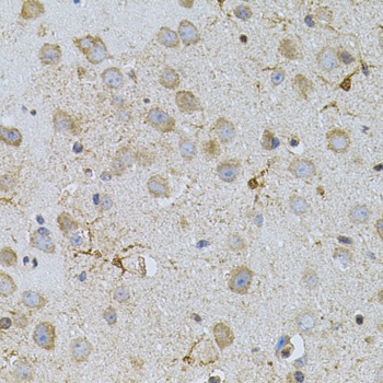
Immunohistochemistry analysis of paraffin-embedded Mouse brain using NAT8 Rabbit pAb (CAB7759) (40x lens). Microwave antigen retrieval performed with 0.01M PBS Buffer (pH 7.2) prior to IHC staining.
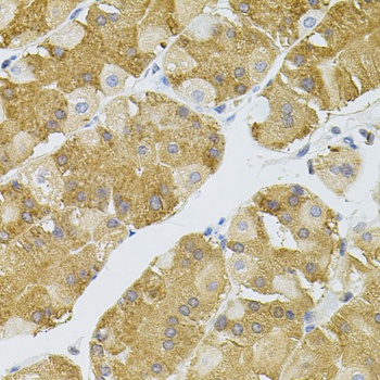
Immunohistochemistry analysis of paraffin-embedded Human stomach using NAT8 Rabbit pAb (CAB7759) (40x lens). Microwave antigen retrieval performed with 0.01M PBS Buffer (pH 7.2) prior to IHC staining.