The NCAPH Antibody (CAB4548) is a high-quality antibody developed for reliable detection and analysis of target proteins. This antibody is produced in rabbits and exhibits high reactivity with human samples, making it suitable for use in Western blot applications. By specifically binding to the Ncaph protein, this antibody enables precise detection and analysis in various cell types, making it ideal for studies in genetics, cell biology, and cancer research.
This antibody is validated for use in WB, IF/ICC, ELISA applications and has demonstrated reactivity against Human samples.
Product Name:
NCAPH Antibody
SKU:
CAB4548
Size:
20μL, 100μL
Reactivity:
Human
Conjugate:
Unconjugated
Immunogen:
Recombinant protein (or fragment).This information is considered to be commercially sensitive.
Recommended starting concentration is 1 μg/mL. Please optimize the concentration based on your specific assay requirements.
Synonyms:
CAPH, BRRN1, CAP-H, MCPH23, NCAPH
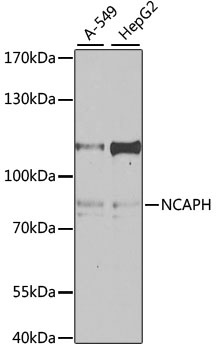
Positive Sample:
A-549, HepG2
Cellular Localization:
Chromosome, Cytoplasm, Nucleus.
Calculated MW:
83kDa
Observed MW:
85kDa
This gene encodes a member of the barr gene family and a regulatory subunit of the condensin complex. This complex is required for the conversion of interphase chromatin into condensed chromosomes. The protein encoded by this gene is associated with mitotic chromosomes, except during the early phase of chromosome condensation. During interphase, the protein has a distinct punctate nucleolar localization. Alternatively spliced transcript variants encoding different proteins have been described.
Purification Method
Affinity purification
Gene ID
23397
RRID
AB_2765747
Buffer Information
Store at -20℃. Avoid freeze / thaw cycles. Buffer: PBS containing 50% glycerol, preserved with proclin300 or sodium azide, pH 7.3.
Western blot analysis of various lysates using NCAPH Rabbit pAb (CAB4548) at 1:1000 dilution. Secondary antibody: HRP-conjugated Goat anti-Rabbit IgG (H+L) (CABS014) at 1:10000 dilution. Lysates/proteins: 25μg per lane. Blocking buffer: 3% nonfat dry milk in TBST. Detection: ECL Enhanced Kit (AbGn00021). Exposure time: 60s.
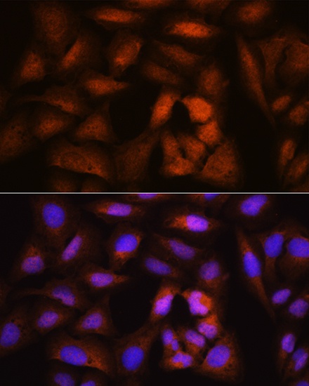
Immunofluorescence analysis of U2OS cells using NCAPH Rabbit pAb (CAB4548) at dilution of 1:100. Secondary antibody: Cy3-conjugated Goat anti-Rabbit IgG (H+L) (CABS007) at 1:500 dilution. Blue: DAPI for nuclear staining.