The NCF4 Monoclonal Antibody (CAB0935) is a high-quality antibody developed for reliable detection and analysis of target proteins. This antibody, raised in rabbits, is known for its high reactivity with human samples and is validated for use in Western blot applications.NCF4 is a key component of the phagocytic NADPH oxidase complex, essential for the production of reactive oxygen species in response to infections and inflammation. Dysregulation of NCF4 has been implicated in various inflammatory disorders, making it an important target for research in immunology and infectious diseases.
This antibody is validated for use in WB, IHC-P, IP, ELISA applications and has demonstrated reactivity against Human samples.
Product Name:
NCF4 Monoclonal Antibody
SKU:
CAB0935
Size:
20μL, 100μL
Reactivity:
Human
Clone Number:
ARC2553
Conjugate:
Unconjugated
Immunogen:
Synthetic peptide. This information is considered to be commercially sensitive.
The protein encoded by this gene is a cytosolic regulatory component of the superoxide-producing phagocyte NADPH-oxidase, a multicomponent enzyme system important for host defense. This protein is preferentially expressed in cells of myeloid lineage. It interacts primarily with neutrophil cytosolic factor 2 (NCF2/p67-phox) to form a complex with neutrophil cytosolic factor 1 (NCF1/p47-phox), which further interacts with the small G protein RAC1 and translocates to the membrane upon cell stimulation. This complex then activates flavocytochrome b, the membrane-integrated catalytic core of the enzyme system. The PX domain of this protein can bind phospholipid products of the PI(3) kinase, which suggests its role in PI(3) kinase-mediated signaling events. The phosphorylation of this protein was found to negatively regulate the enzyme activity. Alternatively spliced transcript variants encoding distinct isoforms have been observed.
Purification Method
Affinity purification
Gene ID
4689
Buffer Information
Store at -20℃. Avoid freeze / thaw cycles. Buffer: PBS containing 50% glycerol and 0.05% BSA, preserved with proclin300 or sodium azide, pH 7.3.
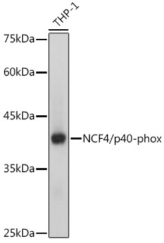
Western blot analysis of lysates from THP-1 cells, using NCF4/p40-phox Rabbit mAb (CAB0935) at 1:1000 dilution. Secondary antibody: HRP-conjugated Goat anti-Rabbit IgG (H+L) (CABS014) at 1:10000 dilution. Lysates/proteins: 25μg per lane. Blocking buffer: 3% nonfat dry milk in TBST. Detection: ECL Basic Kit (AbGn00020). Exposure time: 1s.
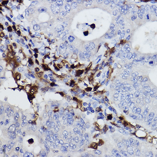
Immunohistochemistry analysis of paraffin-embedded Human colon carcinoma using NCF4/p40-phox Rabbit mAb (CAB0935) at dilution of 1:100 (40x lens). High pressure antigen retrieval performed with 0.01M Citrate buffer (pH 6.0) prior to IHC staining.