The [KO Validated] NCOA4 Antibody (CAB5695) is a high-quality antibody developed for reliable detection and analysis of target proteins. This gene encodes an androgen receptor coactivator. The encoded protein interacts with the androgen receptor in a ligand-dependent manner to enhance its transcriptional activity. Chromosomal translocations between this gene and the ret tyrosine kinase gene, also located on chromosome 10, have been associated with papillary thyroid carcinoma. Alternatively spliced transcript variants have been described. Pseudogenes are present on chromosomes 4, 5, 10, and 14.
This antibody is validated for use in WB, IHC-P, IF/ICC, ELISA applications and has demonstrated reactivity against Human, Mouse, Rat samples.
Product Name:
[KO Validated] NCOA4 Antibody
SKU:
CAB5695
Size:
100μL, 20μL
Reactivity:
Human, Mouse, Rat
Conjugate:
Unconjugated
Immunogen:
Recombinant protein (or fragment).This information is considered to be commercially sensitive.
Tested Applications:
WBIHC-PIF/ICCELISA
Recommended Dilution:
WB
1:500 - 1:1000
IHC-P
1:50 - 1:200
IF/ICC
1:50 - 1:200
ELISA
Recommended starting concentration is 1 μg/mL. Please optimize the concentration based on your specific assay requirements.
Synonyms:
RFG, ELE1, PTC3, ARA70, NCOA4
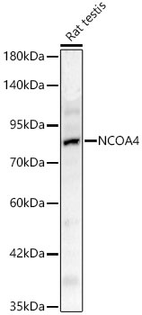
Positive Sample:
Rat testis
Cellular Localization:
Nucleus.
Calculated MW:
70kDa
Observed MW:
80kDa
This gene encodes an androgen receptor coactivator. The encoded protein interacts with the androgen receptor in a ligand-dependent manner to enhance its transcriptional activity. Chromosomal translocations between this gene and the ret tyrosine kinase gene, also located on chromosome 10, have been associated with papillary thyroid carcinoma. Alternatively spliced transcript variants have been described. Pseudogenes are present on chromosomes 4, 5, 10, and 14.
Purification Method
Affinity purification
Gene ID
8031
RRID
AB_2766454
Buffer Information
Store at -20℃. Avoid freeze / thaw cycles. Buffer: PBS with 0.09% Sodium azide,50% glycerol,pH7.3.
Western blot analysis of lysates from Rat testis using NCOA4 Rabbit pAb (CAB5695) at 1:1000 dilution. Secondary antibody: HRP-conjugated Goat anti-Rabbit IgG (H+L) (AS014) at 1:10000 dilution. Lysates/proteins: 25 μg per lane. Blocking buffer: 3% nonfat dry milk in TBST. Detection: ECL Basic Kit (AbGn00020). Exposure time: 45s.
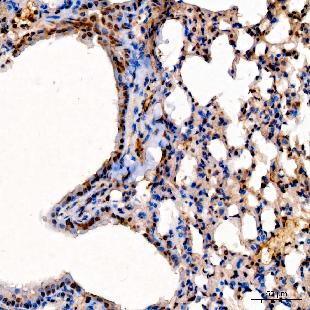
Immunohistochemistry analysis of paraffin-embedded Mouse lung tissue using NCOA4 Rabbit pAb (CAB5695) at a dilution of 1:50 (40x lens). High pressure antigen retrieval performed with 0.01M Citrate buffer (pH 6.0) prior to IHC staining.
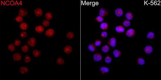
Immunofluorescence analysis of K-562 cells using NCOA4 Rabbit pAb(CAB5695) at a dilution of 1:100 (40x lens). Secondary antibody:Cy3 Goat Anti-Rabbit IgG (H+L)(AS007) at 1:500 dilution. Blue: DAPI for nuclear staining.
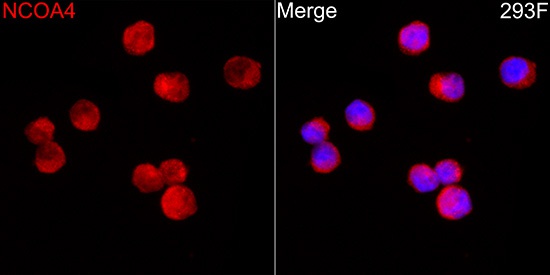
Immunofluorescence analysis of 293F cells using NCOA4 Rabbit pAb(CAB5695) at a dilution of 1:100 (40x lens). Secondary antibody:Cy3 Goat Anti-Rabbit IgG (H+L)(AS007) at 1:500 dilution. Blue: DAPI for nuclear staining.