The NDUFA2 Antibody (CAB8136) is a high-quality antibody developed for reliable detection and analysis of target proteins. This antibody, generated in rabbits, is highly specific to NDUFA2 in human samples and has been validated for use in Western blot applications. By binding to the NDUFA2 protein, this antibody enables accurate detection and analysis in a variety of cell types, making it an essential asset for research in mitochondrial biology and related fields.NDUFA2 is a key component of mitochondrial complex I, essential for the generation of ATP through oxidative phosphorylation. Dysregulation of NDUFA2 has been implicated in various diseases, including neurodegenerative disorders and metabolic syndromes.
This antibody is validated for use in WB, IF/ICC, ELISA applications and has demonstrated reactivity against Human, Mouse, Rat samples.
Product Name:
NDUFA2 Antibody
SKU:
CAB8136
Size:
20μL, 100μL
Reactivity:
Human, Mouse, Rat
Conjugate:
Unconjugated
Immunogen:
Recombinant protein (or fragment).This information is considered to be commercially sensitive.
The encoded protein is a subunit of the hydrophobic protein fraction of the NADH:ubiquinone oxidoreductase (complex 1), the first enzyme complex in the electron transport chain located in the inner mitochondrial membrane, and may be involved in regulating complex I activity or its assembly via assistance in redox processes. Mutations in this gene are associated with Leigh syndrome, an early-onset progressive neurodegenerative disorder. Alternative splicing results in multiple transcript variants.
Purification Method
Affinity purification
Gene ID
4695
RRID
AB_2770545
Buffer Information
Store at -20℃. Avoid freeze / thaw cycles. Buffer: PBS containing 50% glycerol, preserved with proclin300 or sodium azide, pH 7.3.
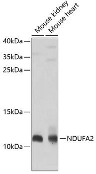
Western blot analysis of various lysates using NDUFA2 Rabbit pAb (CAB8136) at 1:1000 dilution. Secondary antibody: HRP-conjugated Goat anti-Rabbit IgG (H+L) (CABS014) at 1:10000 dilution. Lysates/proteins: 25μg per lane. Blocking buffer: 3% nonfat dry milk in TBST. Detection: ECL Basic Kit (AbGn00020). Exposure time: 30s.
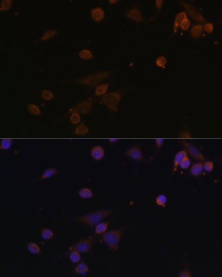
Immunofluorescence analysis of NIH/3T3 cells using NDUFA2 Rabbit pAb (CAB8136) at dilution of 1:100. Secondary antibody: Cy3-conjugated Goat anti-Rabbit IgG (H+L) (CABS007) at 1:500 dilution. Blue: DAPI for nuclear staining.