The NDUFAF2 Antibody (CAB14296) is a high-quality antibody developed for reliable detection and analysis of target proteins. This antibody, generated in rabbits, is highly specific for human samples and has been validated for use in Western blot applications. By binding to the NDUFAF2 protein, researchers can easily detect and analyze its expression in a variety of cell types, making it ideal for studies in mitochondrial biology and related fields.NDUFAF2 plays a crucial role in the assembly of mitochondrial complex I, the largest multimeric enzyme complex in the mitochondrial electron transport chain.
This antibody is validated for use in WB, IF/ICC, ELISA applications and has demonstrated reactivity against Human, Mouse, Rat samples.
Product Name:
NDUFAF2 Antibody
SKU:
CAB14296
Size:
20μL, 100μL
Reactivity:
Human, Mouse, Rat
Conjugate:
Unconjugated
Immunogen:
Recombinant protein (or fragment).This information is considered to be commercially sensitive.
Recommended starting concentration is 1 μg/mL. Please optimize the concentration based on your specific assay requirements.
Synonyms:
MMTN, B17.2L, MC1DN10, mimitin, NDUFA12L, NDUFAF2
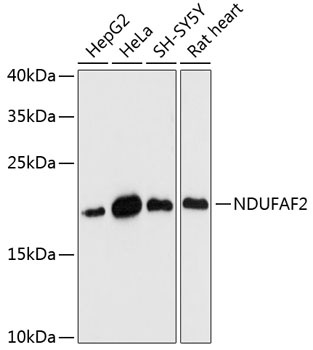
Positive Sample:
HepG2, HeLa, SH-SY5Y, Rat heart
Cellular Localization:
Mitochondrion.
Calculated MW:
20kDa
Observed MW:
20kDa
NADH:ubiquinone oxidoreductase (complex I) catalyzes the transfer of electrons from NADH to ubiquinone (coenzyme Q) in the first step of the mitochondrial respiratory chain, resulting in the translocation of protons across the inner mitochondrial membrane. This gene encodes a complex I assembly factor. Mutations in this gene cause progressive encephalopathy resulting from mitochondrial complex I deficiency.
Purification Method
Affinity purification
Gene ID
91942
RRID
AB_2761158
Buffer Information
Store at -20℃. Avoid freeze / thaw cycles. Buffer: PBS with 0.01% thimerosal,50% glycerol,pH7.3.
Western blot analysis of various lysates using NDUFAF2 Rabbit pAb (CAB14296) at 1:3000 dilution. Secondary antibody: HRP-conjugated Goat anti-Rabbit IgG (H+L) (CABS014) at 1:10000 dilution. Lysates/proteins: 25μg per lane. Blocking buffer: 3% nonfat dry milk in TBST. Detection: ECL Basic Kit (AbGn00020). Exposure time: 90s.
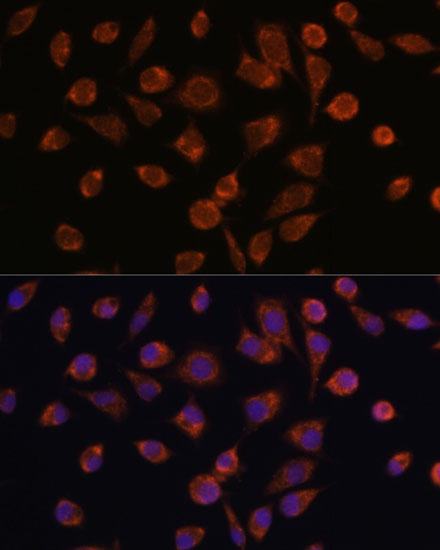
Immunofluorescence analysis of L929 cells using NDUFAF2 Rabbit pAb (CAB14296) at dilution of 1:100. Secondary antibody: Cy3-conjugated Goat anti-Rabbit IgG (H+L) (CABS007) at 1:500 dilution. Blue: DAPI for nuclear staining.