The NDUFS1 Antibody (CAB16926) is a high-quality antibody developed for reliable detection and analysis of target proteins. This antibody, raised in rabbits, exhibits high reactivity with human samples and has been validated for use in Western blot applications. By binding to the NDUFS1 protein, it allows for the detection and analysis of NDUFS1 in various cell types, making it an essential tool for studies in metabolism, bioenergetics, and mitochondrial dysfunction.NDUFS1 is essential for the proper functioning of complex I in the electron transport chain, which is responsible for generating the majority of ATP in cells.
This antibody is validated for use in WB, IHC-P, IF/ICC, ELISA applications and has demonstrated reactivity against Human, Mouse, Rat samples.
Product Name:
NDUFS1 Antibody
SKU:
CAB16926
Size:
20μL, 100μL
Reactivity:
Human, Mouse, Rat
Immunogen:
Recombinant protein (or fragment).This information is considered to be commercially sensitive.
The protein encoded by this gene belongs to the complex I 75 kDa subunit family. Mammalian complex I is composed of 45 different subunits. It locates at the mitochondrial inner membrane. This protein has NADH dehydrogenase activity and oxidoreductase activity. It transfers electrons from NADH to the respiratory chain. The immediate electron acceptor for the enzyme is believed to be ubiquinone. This protein is the largest subunit of complex I and it is a component of the iron-sulfur (IP) fragment of the enzyme. It may form part of the active site crevice where NADH is oxidized. Mutations in this gene are associated with complex I deficiency. Several transcript variants encoding different isoforms have been found for this gene.
Purification Method
Affinity purification
Gene ID
4719
RRID
AB_2770551
Buffer Information
Store at -20℃. Avoid freeze / thaw cycles. Buffer: PBS containing 50% glycerol, preserved with proclin300 or sodium azide, pH 7.3.
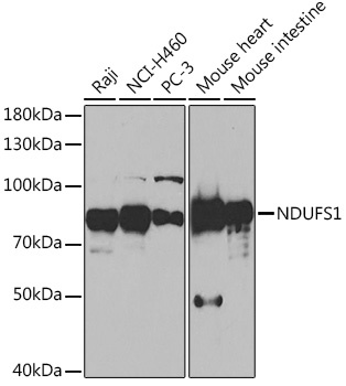
Western blot analysis of various lysates using NDUFS1 Rabbit pAb (CAB16926) at 1:1000 dilution. Secondary antibody: HRP-conjugated Goat anti-Rabbit IgG (H+L) (CABS014) at 1:10000 dilution. Lysates/proteins: 25μg per lane. Blocking buffer: 3% nonfat dry milk in TBST. Detection: ECL Basic Kit (AbGn00020). Exposure time: 90s.
Immunohistochemistry analysis of paraffin-embedded Mouse kidney using NDUFS1 Rabbit pAb (CAB16926) at dilution of 1:50 (40x lens). High pressure antigen retrieval performed with 0.01M Citrate buffer (pH 6.0) prior to IHC staining.
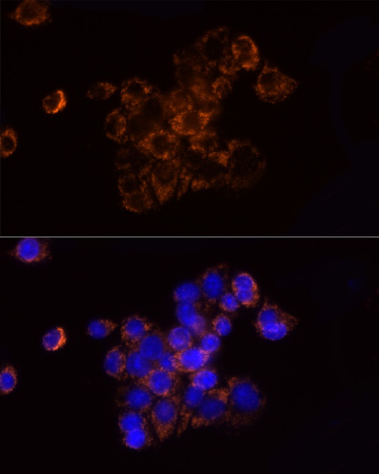
Immunofluorescence analysis of HepG2 cells using NDUFS1 Rabbit pAb (CAB16926) at dilution of 1:50 (40x lens). Secondary antibody: Cy3-conjugated Goat anti-Rabbit IgG (H+L) (CABS007) at 1:500 dilution. Blue: DAPI for nuclear staining.
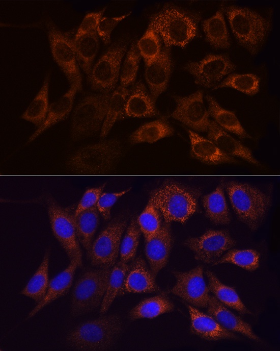
Immunofluorescence analysis of NIH/3T3 cells using NDUFS1 Rabbit pAb (CAB16926) at dilution of 1:50 (40x lens). Secondary antibody: Cy3-conjugated Goat anti-Rabbit IgG (H+L) (CABS007) at 1:500 dilution. Blue: DAPI for nuclear staining.