The NDUFV2 Antibody (CAB7442) is a high-quality antibody developed for reliable detection and analysis of target proteins. This antibody, generated in rabbits, exhibits high reactivity with human samples and is suitable for use in Western blot analyses. By targeting the NDUFV2 protein, researchers can accurately detect and analyze its expression in various cell types, making it an excellent tool for studies in mitochondrial biology and related fields.The NDUFV2 protein is essential for the function of complex I in the electron transport chain, which is crucial for ATP production and cellular energy metabolism.
This antibody is validated for use in WB, ELISA applications and has demonstrated reactivity against Human, Mouse, Rat samples.
Product Name:
NDUFV2 Antibody
SKU:
CAB7442
Size:
20μL, 100μL
Reactivity:
Human, Mouse, Rat
Conjugate:
Unconjugated
Immunogen:
Recombinant protein (or fragment).This information is considered to be commercially sensitive.
The NADH-ubiquinone oxidoreductase complex (complex I) of the mitochondrial respiratory chain catalyzes the transfer of electrons from NADH to ubiquinone, and consists of at least 43 subunits. The complex is located in the inner mitochondrial membrane. This gene encodes the 24 kDa subunit of complex I, and is involved in electron transfer. Mutations in this gene are implicated in Parkinson's disease, bipolar disorder, schizophrenia, and have been found in one case of early onset hypertrophic cardiomyopathy and encephalopathy. A non-transcribed pseudogene of this locus is found on chromosome 19.
Purification Method
Affinity purification
Gene ID
4729
RRID
AB_2767973
Buffer Information
Store at -20℃. Avoid freeze / thaw cycles. Buffer: PBS containing 50% glycerol, preserved with proclin300 or sodium azide, pH 7.3.
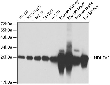
Western blot analysis of various lysates using NDUFV2 Rabbit pAb (CAB7442) at 1:1000 dilution. Secondary antibody: HRP-conjugated Goat anti-Rabbit IgG (H+L) (CABS014) at 1:10000 dilution. Lysates/proteins: 25μg per lane. Blocking buffer: 3% nonfat dry milk in TBST. Detection: ECL Basic Kit (AbGn00020). Exposure time: 30s.