The NEK8 Antibody (CAB0984) is a high-quality antibody developed for reliable detection and analysis of target proteins. This gene encodes a member of the serine/threionine protein kinase family related to NIMA (never in mitosis, gene A) of Aspergillus nidulans. The encoded protein may play a role in cell cycle progression from G2 to M phase. Mutations in the related mouse gene are associated with a disease phenotype that closely parallels the juvenile autosomal recessive form of polycystic kidney disease in humans.
This antibody is validated for use in WB, IF/ICC, ELISA applications and has demonstrated reactivity against Human, Mouse, Rat samples.
Product Name:
NEK8 Antibody
SKU:
CAB0984
Size:
100μL, 20μL
Reactivity:
Human, Mouse, Rat
Conjugate:
Unconjugated
Immunogen:
Recombinant protein (or fragment).This information is considered to be commercially sensitive.
Tested Applications:
WBIF/ICCELISA
Recommended Dilution:
WB
1:100 - 1:500
IF/ICC
1:100 - 1:500
ELISA
Recommended starting concentration is 1 μg/mL. Please optimize the concentration based on your specific assay requirements.
Synonyms:
JCK, NPHP9, RHPD2, NEK12A, NEK8
Positive Sample:
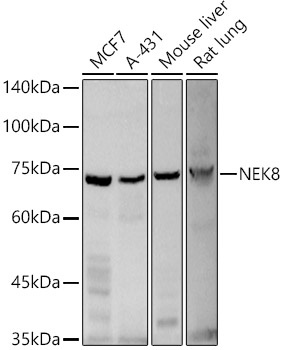
MCF7, A-431, Mouse liver, Rat lung,
Cellular Localization:
Cell Projection, Cytoplasm, Cilium, Cytoskeleton.
Calculated MW:
75kDa
Observed MW:
70kDa/
This gene encodes a member of the serine/threionine protein kinase family related to NIMA (never in mitosis, gene A) of Aspergillus nidulans. The encoded protein may play a role in cell cycle progression from G2 to M phase. Mutations in the related mouse gene are associated with a disease phenotype that closely parallels the juvenile autosomal recessive form of polycystic kidney disease in humans.
Purification Method
Affinity purification
Gene ID
284086
RRID
AB_2757503
Buffer Information
Store at -20℃. Avoid freeze / thaw cycles. Buffer: PBS containing 50% glycerol, preserved with proclin300 or sodium azide, pH 7.3.
Western blot analysis of various lysates using NEK8 Rabbit pAb (CAB0984) at 1:500 dilution. Secondary antibody: HRP-conjugated Goat anti-Rabbit IgG (H+L) (AS014) at 1:10000 dilution. Lysates / proteins: 25 μg per lane. Blocking buffer: 3 % nonfat dry milk in TBST. Detection: ECL Basic Kit (AbGn00020). Exposure time: 180s.
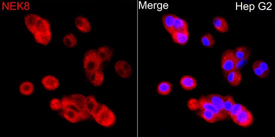
Immunofluorescence analysis of HepG2 cells using NEK8 Rabbit pAb (CAB0984) at dilution of 1:500 (40x lens). Secondary antibody: Cy3-conjugated Goat anti-Rabbit IgG (H+L) (AS007) at 1:500 dilution. Blue: DAPI for nuclear staining.