The NPHP1 Antibody (CAB6674) is a high-quality antibody developed for reliable detection and analysis of target proteins. This antibody, generated in rabbits, exhibits high specificity and reactivity with human samples, making it an ideal choice for immunofluorescence and immunohistochemistry applications.Nephrocystin-1 is a key player in the development and maintenance of primary cilia, cellular appendages with crucial roles in signaling pathways and cell cycle regulation.
This antibody is validated for use in WB, IHC-P, IF/ICC, ELISA applications and has demonstrated reactivity against Human, Mouse, Rat samples.
Product Name:
NPHP1 Antibody
SKU:
CAB6674
Size:
20μL, 100μL
Reactivity:
Human, Mouse, Rat
Conjugate:
Unconjugated
Immunogen:
Recombinant protein (or fragment).This information is considered to be commercially sensitive.
This gene encodes a protein with src homology domain 3 (SH3) patterns. This protein interacts with Crk-associated substrate, and it appears to function in the control of cell division, as well as in cell-cell and cell-matrix adhesion signaling, likely as part of a multifunctional complex localized in actin- and microtubule-based structures. Mutations in this gene cause familial juvenile nephronophthisis type 1, a kidney disorder involving both tubules and glomeruli. Defects in this gene are also associated with Senior-Loken syndrome type 1, also referred to as juvenile nephronophthisis with Leber amaurosis, which is characterized by kidney and eye disease, and with Joubert syndrome type 4, which is characterized by cerebellar ataxia, oculomotor apraxia, psychomotor delay and neonatal breathing abnormalities, sometimes including retinal dystrophy and renal disease. Multiple transcript variants encoding different isoforms have been found for this gene.
Purification Method
Affinity purification
Gene ID
4867
RRID
AB_2767259
Buffer Information
Store at -20℃. Avoid freeze / thaw cycles. Buffer: PBS containing 50% glycerol, preserved with proclin300 or sodium azide, pH 7.3.
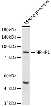
Western blot analysis of lysates from Mouse pancreas, using NPHP1 Rabbit pAb (CAB6674) at 1:1000 dilution. Secondary antibody: HRP-conjugated Goat anti-Rabbit IgG (H+L) (CABS014) at 1:10000 dilution. Lysates/proteins: 25μg per lane. Blocking buffer: 3% nonfat dry milk in TBST. Detection: ECL Basic Kit (AbGn00020). Exposure time: 180s.
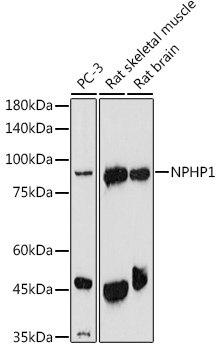
Western blot analysis of various lysates using NPHP1 Rabbit pAb (CAB6674) at 1:1000 dilution. Secondary antibody: HRP-conjugated Goat anti-Rabbit IgG (H+L) (CABS014) at 1:10000 dilution. Lysates/proteins: 25μg per lane. Blocking buffer: 3% nonfat dry milk in TBST. Detection: ECL Enhanced Kit (AbGn00021). Exposure time: 180s.
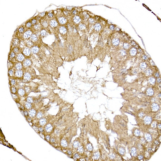
Immunohistochemistry analysis of paraffin-embedded Rat testis using NPHP1 Rabbit pAb (CAB6674) at dilution of 1:50 (40x lens). High pressure antigen retrieval performed with 0.01M Citrate buffer (pH 6.0) prior to IHC staining.
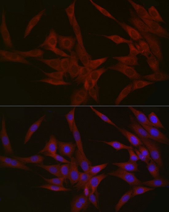
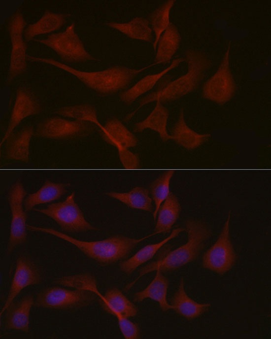
Immunofluorescence analysis of NIH/3T3 cells using NPHP1 Rabbit pAb (CAB6674) at dilution of 1:50 (40x lens). Secondary antibody: Cy3-conjugated Goat anti-Rabbit IgG (H+L) (CABS007) at 1:500 dilution. Blue: DAPI for nuclear staining.
Immunofluorescence analysis of U2OS cells using NPHP1 Rabbit pAb (CAB6674) at dilution of 1:50 (40x lens). Secondary antibody: Cy3-conjugated Goat anti-Rabbit IgG (H+L) (CABS007) at 1:500 dilution. Blue: DAPI for nuclear staining.


 at 1:10000 dilution. Lysates/proteins: 25ug per lane. Blocking buffer: 3% nonfat dry milk in TBST. Detection: ECL Basic Kit. Exposure time: 180s.")
 at 1:10000 dilution. Lysates/proteins: 25ug per lane. Blocking buffer: 3% nonfat dry milk in TBST. Detection: ECL Basic Kit. Exposure time: 90s.")


