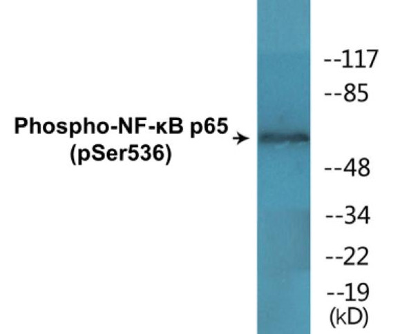
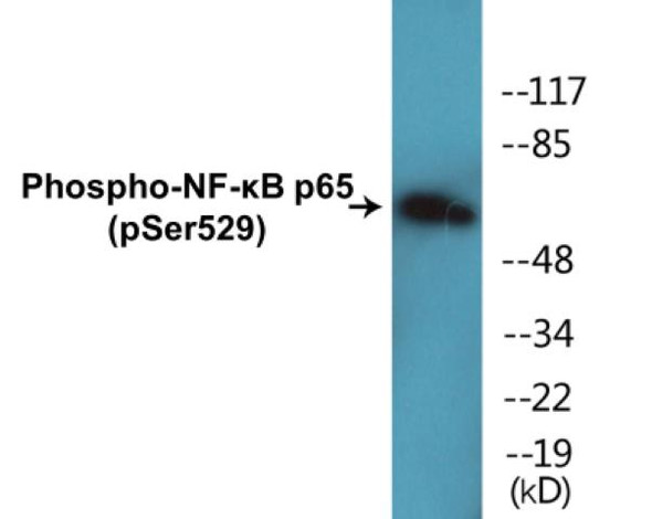
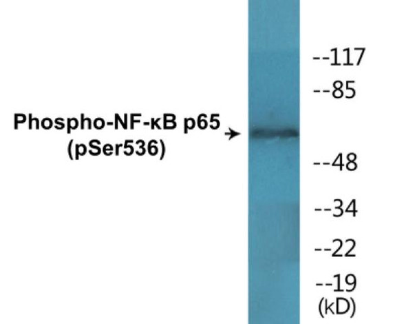
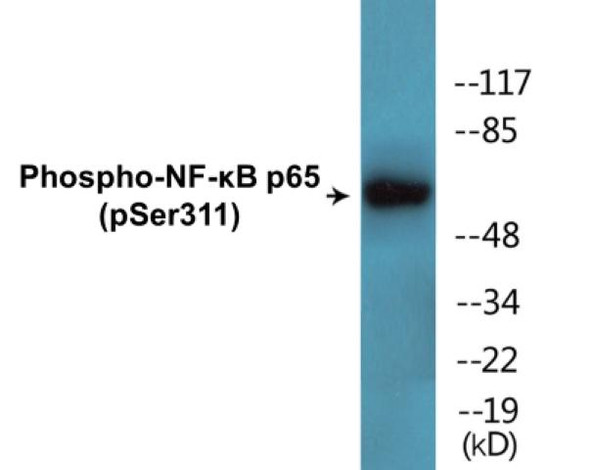
Description
NF-kB p65 (Phospho-Ser536) Cell-Based ELISA Kit
The NF-kB p65 (Phospho-Ser536) Cell-Based ELISA Kit is a convenient, lysate- free, high throughput and sensitive assay kit that can monitor NF-kB p65 phosphorylation and expression profile in cells. The kit can be used for measuring the relative amounts of phosphorylated NF-kB p65 in cultured cells as well as screening for the effects that various treatments, inhibitors (ie. siRNA or chemicals), or activators have on NF-kB p65 phosphorylation.
How does our NF-kB p65 (Phospho-Ser536) Fluorometric Cell-Based ELISA Kit work?
Qualitative determination of NF-kB p65 (Phospho-Ser536) concentration is achieved by an indirect ELISA format. In essence, NF-kB p65 (Phospho-Ser536) is captured by NF-kB p65 (Phospho-Ser536)-specific primary (1°) antibodies while Dye 1-conjugated and Dye 2-conjugated secondary (2°) antibodies bind the Fc region of the 1° antibody. Through this binding, the dye conjugated to the 2° antibody can emit light at a certain wavelength given proper excitation, hence allowing for a fluorometric detection method. Due to the qualitative nature of the Cell-Based ELISA, multiple normalization methods are needed:
| 1. | A monoclonal antibody specific for human GAPDH is included to serve as an internal positive control in normalizing the target RFU values. |
| 2. | An antibody against the nonphosphorylated counterpart of NF-kB p65 (Phospho-Ser536) is also provided for normalization purposes. The RFU values obtained for non-phosphorylated NF-kB p65 can be used to normalize the RFU value for phosphorylated NF-kB p65. |
NF-kB p65 (Phospho-Ser536) Fluorometric Cell-Based ELISA Kit -Information
| Product Name: | NF-kB p65 (Phospho-Ser536)Fluorometric Cell-Based ELISA Kit |
| Product Code/SKU: | FBCAB00075 |
| Description: | The NF-kB p65 (Phospho-Ser536) Fluorometric Cell-Based Phospho ELISA Kit is a convenient, lysate-free, high throughput and sensitive assay kit that can monitor NF-kB p65 (Phospho-Ser536) protein phosphorylation and expression profile in cells. The kit can be used for measuring the relative amounts of phosphorylated NF-kB p65 (Phospho-Ser536) in cultured cells as well as screening for the effects that various treatments, inhibitors (ie. siRNA or chemicals, or activators have on RELA phosphorylation. |
| Dynamic Range: | > 5000 Cells |
| Detection Method: | Fluorometric |
| Storage/Stability: | 4°C/6 Months |
| Reactivity: | Human, Mouse, Rat |
| Assay Type: | Cell-Based ELISA |
| Database Links: | Gene ID: 5970, UniProt ID: Q04206, OMIM #: 164014, Unigene #: Hs.502875 |
| Format: | Two 96-Well Plates |
| NCBI Gene Symbol: | RELA |
| Sub Type: | Phospho |
| Target Name: | Phospho-NF-?B p65 (Ser536) |
Kit Principle
Figure: Schematic representation of Assay Genie Cell-Based Fluorometric ELISA principle
Kit components | Quantity |
| 96-Well Black Cell CultureClear-Bottom Microplate | 2 plates |
| 10X TBS | 24 ml |
| Quenching Buffer | 24 ml |
| Blocking Buffer | 50 ml |
| 15X Wash Buffer | 50 ml |
| Primary Antibody Diluent | 12 ml |
| 100x Anti-Phospho Target Antibody | 60 µl |
| 100x Anti-Target Antibody | 60 µl |
| Anti-GAPDH Antibody | 110 µl |
| Dye-1 Conjugated Anti-Rabbit IgG Antibody | 6 ml |
| Dye-2 Conjugated Anti-Mouse IgG Antibody | 6 ml |
| Adhesive Plate Seals | 2 seals |
Additional equipment and materials required
The following materials and/or equipment are NOT provided in this kit but are necessary to successfully conduct the experiment:
- Fluorescent plate reader with two channels at Ex/Em: 651/667 and 495/521
- Micropipettes capable of measuring volumes from 1 µl to 1 ml
- Deionized or sterile water (ddH2O)
- 37% formaldehyde (Sigma Cat# F-8775) or formaldehyde from other sources
- Squirt bottle, manifold dispenser, multichannel pipette reservoir or automated microplate washer
- Graph paper or computer software capable of generating or displaying logarithmic functions
- Absorbent papers or vacuum aspirator
- Test tubes or microfuge tubes capable of storing ≥1 ml
- Poly-L-Lysine (Sigma Cat# P4832 for suspension cells)
- Orbital shaker (optional)
Kit Protocol
This is a summarized version of the kit protocol. Please view the technical manual of this kit for information on sample preparation, reagent preparation and plate lay out.
| 1. | Seed 200 µl of desired cell concentration in culture medium into each well of the 96-well plates. For suspension cells and loosely attached cells, coat the plates with 100 µl of 10 µg/ml Poly-L-Lysine (not included) to each well of a 96-well plate for 30 minutes at 37°C prior to adding cells. |
| 2. | Incubate the cells for overnight at 37°C, 5% CO2. |
| 3. | Treat the cells as desired. |
| 4. | Remove the cell culture medium and rinse with 200 µl of 1x TBS, twice. |
| 5. | Fix the cells by incubating with 100 µl of Fixing Solution for 20 minutes at room temperature. The 4% formaldehyde is used for adherent cells and 8% formaldehyde is used for suspension cells and loosely attached cells. |
| 6. | Remove the Fixing Solution and wash the plate 3 times with 200 µl 1x Wash Buffer for 3 minutes. The plate can be stored at 4°C for a week. |
| 7. | Add 100 µl of Quenching Buffer and incubate for 20 minutes at room temperature. |
| 8. | Wash the plate 3 times with 1x Wash Buffer for 3 minutes each time. |
| 9. | Dispense 200 µl of Blocking Buffer and incubate for 1 hour at room temperature. |
| 10. | Wash 3 times with 200 µl of 1x Wash Buffer for 3 minutes each time. |
| 11. | Add 50 µl of Primary Antibody Mixture P to corresponding wells for NF-kB p65 (Phospho-Ser536) detection. Add 50 µl of Primary Antibody Mixture NP to the corresponding wells for total NF-kB p65 detection. Cover the plate with parafilm and incubate for 16 hours (overnight) at 4°C. If the target expression is known to be high, incubate for 2 hours at room temperature. |
| 12. | Wash 3 times with 200 µl of 1x Wash Buffer for 3 minutes each time. |
| 13. | Add 50 ul of Secondary Antibody Mixture to corresponding wells and incubate for 1.5 hours at room temperature in the dark. |
| 14. | Wash 3 times with 200 µl of 1x Wash Buffer for 3 minutes each time. |
| 15. | Read the plate(s) at Ex/Em: 651/667 (Dye 1) and 495/521 (Dye 2). Shield plates from direct light exposure. |
| 16. | Wash 3 times with 200 µl of 1x Wash Buffer for 5 minutes each time. |
NF-kB p65 (Phospho-Ser536) - Protein Information
| UniProt Protein Function: | NFkB-p65: a subunit of NF-kappa-B transcription complex, which plays a crucial role in inflammatory and immune responses. The inhibitory effect of I-kappa-B upon NF-kappa-B in the cytoplasm is exerted primarily through the interaction with p65. P65 shows a weak DNA-binding site which could contribute directly to DNA binding in the NF-kappa-B complex. There are five NFkB proteins in mammals (RelA/NFkB-p65, RelB, c-Rel, NF-_B1/NFkB-p105, and NF-_B2/NFkB-p100). They form a variety of homodimers and heterodimers, each of which activates its own characteristic set of genes. Three splice-variant isoforms have been identified. |
| UniProt Protein Details: | Protein type:DNA-binding; Nuclear receptor co-regulator; Transcription factor Chromosomal Location of Human Ortholog: 11q13.1 Cellular Component: cytoplasm; cytosol; nuclear chromatin; nucleoplasm; nucleus; transcription factor complex Molecular Function:actinin binding; chromatin binding; chromatin DNA binding; DNA binding; histone deacetylase binding; identical protein binding; NF-kappaB binding; phosphate binding; protein binding; protein heterodimerization activity; protein homodimerization activity; protein kinase binding; protein N-terminus binding; RNA polymerase II transcription factor activity, enhancer binding; transcription activator binding; transcription factor activity; transcription factor binding; ubiquitin protein ligase binding Biological Process: activation of NF-kappaB transcription factor; cytokine and chemokine mediated signaling pathway; inflammatory response; membrane protein intracellular domain proteolysis; negative regulation of apoptosis; negative regulation of transcription from RNA polymerase II promoter; negative regulation of transcription, DNA-dependent; positive regulation of cell proliferation; positive regulation of I-kappaB kinase/NF-kappaB cascade; positive regulation of interferon type I production; positive regulation of transcription from RNA polymerase II promoter; positive regulation of transcription, DNA-dependent; regulation of inflammatory response; response to organic substance; response to UV-B; stimulatory C-type lectin receptor signaling pathway; T cell receptor signaling pathway |
| NCBI Summary: | NF-kappa-B is a ubiquitous transcription factor involved in several biological processes. It is held in the cytoplasm in an inactive state by specific inhibitors. Upon degradation of the inhibitor, NF-kappa-B moves to the nucleus and activates transcription of specific genes. NF-kappa-B is composed of NFKB1 or NFKB2 bound to either REL, RELA, or RELB. The most abundant form of NF-kappa-B is NFKB1 complexed with the product of this gene, RELA. Four transcript variants encoding different isoforms have been found for this gene. [provided by RefSeq, Sep 2011] |
| UniProt Code: | Q04206 |
| NCBI GenInfo Identifier: | 62906901 |
| NCBI Gene ID: | 5970 |
| NCBI Accession: | Q04206.2 |
| UniProt Secondary Accession: | Q04206,Q6GTV1, Q6SLK1, |
| UniProt Related Accession: | Q04206 |
| Molecular Weight: | 59,910 Da |
| NCBI Full Name: | Transcription factor p65 |
| NCBI Synonym Full Names: | RELA proto-oncogene, NF-kB subunit |
| NCBI Official Symbol: | RELA |
| NCBI Official Synonym Symbols: | p65; NFKB3 |
| NCBI Protein Information: | transcription factor p65 |
| UniProt Protein Name: | Transcription factor p65 |
| UniProt Synonym Protein Names: | Nuclear factor NF-kappa-B p65 subunit; Nuclear factor of kappa light polypeptide gene enhancer in B-cells 3 |
| Protein Family: | Proline-rich P65 protein |
| UniProt Gene Name: | RELA |


