The NFATC3 Antibody (CAB6666) is a high-quality antibody developed for reliable detection and analysis of target proteins. This antibody, produced in rabbits, exhibits high specificity for human samples and is validated for use in Western blot applications. By binding to the NFATC3 protein, this antibody enables precise detection and analysis in various cell types, making it an essential asset for studies in immunology and cancer research.NFATC3 is a crucial player in immune responses, serving as a key mediator in the activation of T cells and B cells.
This antibody is validated for use in WB, IHC-P, IF/ICC, ELISA applications and has demonstrated reactivity against Human, Mouse, Rat samples.
Product Name:
NFATC3 Antibody
SKU:
CAB6666
Size:
20μL, 100μL
Reactivity:
Human, Mouse, Rat
Conjugate:
Unconjugated
Immunogen:
Recombinant protein (or fragment).This information is considered to be commercially sensitive.
Recommended starting concentration is 1 μg/mL. Please optimize the concentration based on your specific assay requirements.
Synonyms:
NFAT4, NFATX, NF-AT4c, n339260, NFATC3
Positive Sample:
Jurkat, Mouse thymus
Cellular Localization:
Cytoplasm, Nucleus.
Calculated MW:
116kDa
Observed MW:
150kDa
The product of this gene is a member of the nuclear factors of activated T cells DNA-binding transcription complex. This complex consists of at least two components: a preexisting cytosolic component that translocates to the nucleus upon T cell receptor (TCR) stimulation and an inducible nuclear component. Other members of this family participate to form this complex also. The product of this gene plays a role in the regulation of gene expression in T cells and immature thymocytes. Several transcript variants encoding distinct isoforms have been identified for this gene.
Purification Method
Affinity purification
Gene ID
4775
RRID
AB_2767253
Buffer Information
Store at -20℃. Avoid freeze / thaw cycles. Buffer: PBS containing 50% glycerol, preserved with proclin300 or sodium azide, pH 7.3.
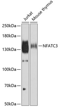
Western blot analysis of various lysates using NFATC3 Rabbit pAb (CAB6666) at 1:500 dilution. Secondary antibody: HRP-conjugated Goat anti-Rabbit IgG (H+L) (CABS014) at 1:10000 dilution. Lysates/proteins: 25μg per lane. Blocking buffer: 3% nonfat dry milk in TBST. Detection: ECL Basic Kit (AbGn00020). Exposure time: 10s.
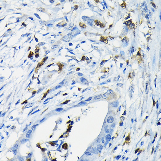
Immunohistochemistry analysis of paraffin-embedded Human colon carcinoma using NFATC3 Rabbit pAb (CAB6666) at dilution of 1:200 (40x lens). High pressure antigen retrieval performed with 0.01M Citrate buffer (pH 6.0) prior to IHC staining.
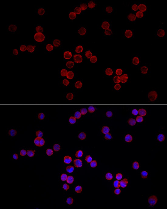
Immunofluorescence analysis of K-562 cells using NFATC3 Rabbit pAb (CAB6666) at dilution of 1:50 (40x lens). Secondary antibody: Cy3-conjugated Goat anti-Rabbit IgG (H+L) (CABS007) at 1:500 dilution. Blue: DAPI for nuclear staining.