The ULBP2 Antibody (CAB8264) is a high-quality antibody developed for reliable detection and analysis of target proteins. This gene encodes a major histocompatibility complex (MHC) class I-related molecule that binds to the NKG2D receptor on natural killer (NK) cells to trigger release of multiple cytokines and chemokines that in turn contribute to the recruitment and activation of NK cells. The encoded protein undergoes further processing to generate the mature protein that is either anchored to membrane via a glycosylphosphatidylinositol moiety, or secreted. Many malignant cells secrete the encoded protein to evade immunosurveillance by NK cells. This gene is located in a cluster of multiple MHC class I-related genes on chromosome 6.
This antibody is validated for use in WB, ELISA applications and has demonstrated reactivity against Human samples.
Product Name:
ULBP2 Antibody
SKU:
CAB8264
Size:
100μL, 20μL
Reactivity:
Human
Conjugate:
Unconjugated
Immunogen:
Synthetic peptide. This information is considered to be commercially sensitive.
Tested Applications:
WBELISA
Recommended Dilution:
WB
1:100 - 1:500
ELISA
Recommended starting concentration is 1 μg/mL. Please optimize the concentration based on your specific assay requirements.
This gene encodes a major histocompatibility complex (MHC) class I-related molecule that binds to the NKG2D receptor on natural killer (NK) cells to trigger release of multiple cytokines and chemokines that in turn contribute to the recruitment and activation of NK cells. The encoded protein undergoes further processing to generate the mature protein that is either anchored to membrane via a glycosylphosphatidylinositol moiety, or secreted. Many malignant cells secrete the encoded protein to evade immunosurveillance by NK cells. This gene is located in a cluster of multiple MHC class I-related genes on chromosome 6.
Purification Method
Affinity purification
Gene ID
80328
RRID
AB_2772809
Buffer Information
Store at -20℃. Avoid freeze / thaw cycles. Buffer: PBS containing 50% glycerol, preserved with proclin300 or sodium azide, pH 7.3.
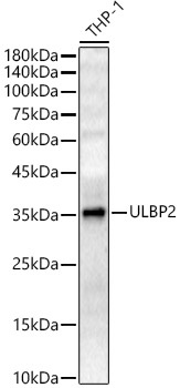
Western blot analysis of lysates from THP-1 cells, using ULBP2 Rabbit pAb (CAB8264) at 1:400 dilution. Secondary antibody: HRP-conjugated Goat anti-Rabbit IgG (H+L) (AS014) at 1:10000 dilution. Lysates/proteins: 25μg per lane. Blocking buffer: 3% nonfat dry milk in TBST. Detection: ECL Enhanced Kit (AbGn00021). Exposure time: 180s.