The NLRC4 Antibody (CAB7382) is a high-quality antibody developed for reliable detection and analysis of target proteins. The antibody, raised in rabbits, is highly reactive with human samples and is validated for use in various applications such as Western blot, immunohistochemistry, and immunofluorescence.NLRC4, also known as NLR family CARD domain-containing protein 4, plays a crucial role in the innate immune system by activating inflammatory responses in the presence of pathogens.
This antibody is validated for use in WB, IF/ICC, ELISA, IF-P applications and has demonstrated reactivity against Human, Mouse, Rat samples.
Product Name:
NLRC4 Antibody
SKU:
CAB7382
Size:
20μL, 100μL
Reactivity:
Human, Mouse, Rat
Conjugate:
Unconjugated
Immunogen:
Recombinant protein (or fragment).This information is considered to be commercially sensitive.
Tested Applications:
WBIF/ICCELISAIF-P
Recommended Dilution:
WB
1:100 - 1:500
IF/ICC
1:50 - 1:200
IF-P
1:50 - 1:200
ELISA
Recommended starting concentration is 1 μg/mL. Please optimize the concentration based on your specific assay requirements.
This gene encodes a member of the caspase recruitment domain-containing NLR family. Family members play essential roles in innate immune response to a wide range of pathogenic organisms, tissue damage and other cellular stresses. Mutations in this gene result in autoinflammation with infantile enterocolitis. Alternative splicing results in multiple transcript variants.
Purification Method
Affinity purification
Gene ID
58484
RRID
AB_2767914
Buffer Information
Store at -20℃. Avoid freeze / thaw cycles. Buffer: PBS containing 50% glycerol, preserved with proclin300 or sodium azide, pH 7.3.
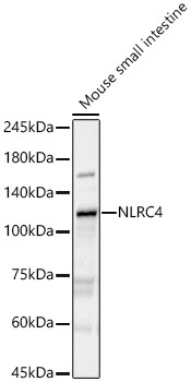
Western blot analysis of lysates from Mouse small intestine, using NLRC4 Rabbit pAb (CAB7382) at 1:400 dilution. Secondary antibody: HRP-conjugated Goat anti-Rabbit IgG (H+L) (CABS014) at 1:10000 dilution. Lysates/proteins: 25μg per lane. Blocking buffer: 3% nonfat dry milk in TBST. Detection: ECL Basic Kit (AbGn00020). Exposure time: 180s.
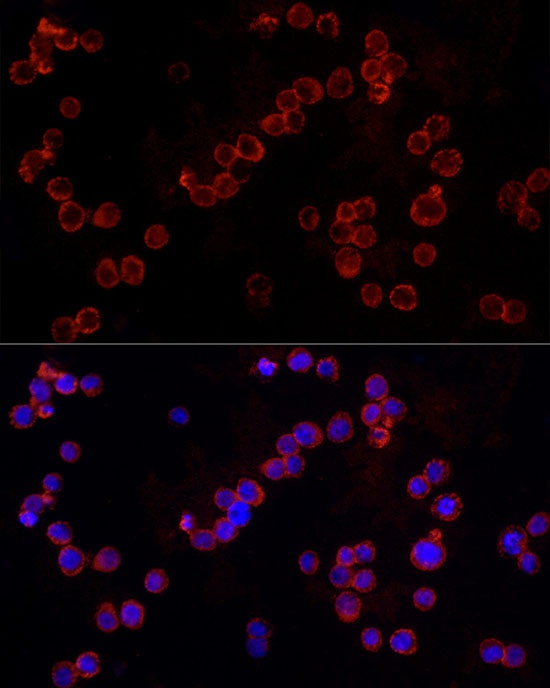
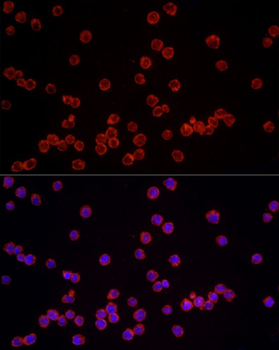
Immunofluorescence analysis of THP-1 cells using NLRC4 Rabbit pAb (CAB7382) at dilution of 1:100 (40x lens). Secondary antibody: Cy3-conjugated Goat anti-Rabbit IgG (H+L) (CABS007) at 1:500 dilution. Blue: DAPI for nuclear staining.
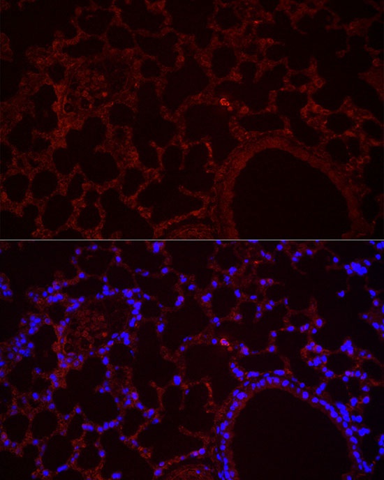
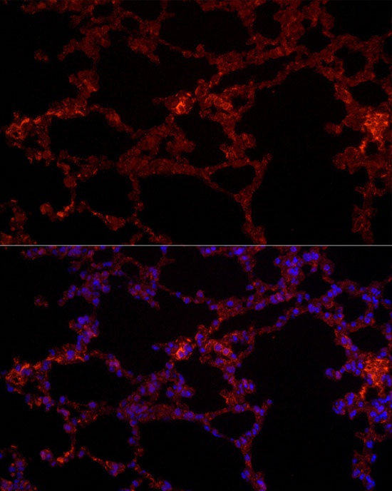
Immunofluorescence analysis of paraffin-embedded mouse lung using NLRC4 Rabbit pAb (CAB7382) at dilution of 1:100 (40x lens). Secondary antibody: Cy3-conjugated Goat anti-Rabbit IgG (H+L) (CABS007) at 1:500 dilution. Blue: DAPI for nuclear staining.
Immunofluorescence analysis of THP-1 cells using NLRC4 Rabbit pAb (CAB7382) at dilution of 1:100 (40x lens). Secondary antibody: Cy3-conjugated Goat anti-Rabbit IgG (H+L) (CABS007) at 1:500 dilution. Blue: DAPI for nuclear staining.
Immunofluorescence analysis of paraffin-embedded mouse lung using NLRC4 Rabbit pAb (CAB7382) at dilution of 1:100 (40x lens). Secondary antibody: Cy3-conjugated Goat anti-Rabbit IgG (H+L) (CABS007) at 1:500 dilution. Blue: DAPI for nuclear staining.