The NLRP3 Polyclonal Antibody (CAB21906) is a high-quality antibody developed for reliable detection and analysis of target proteins. Produced in rabbits, this antibody is highly specific for human samples and has been validated for use in Western blot applications. By targeting the NLRP3 protein, this antibody allows for the detection and analysis of NLRP3 in various cell types, making it an ideal choice for studies in immunology and inflammatory diseases.
This antibody is validated for use in WB, IF/ICC, ELISA applications and has demonstrated reactivity against Human, Mouse, Rat samples.
Product Name:
NLRP3 Polyclonal Antibody
SKU:
CAB21906
Size:
20μL, 100μL
Reactivity:
Human, Mouse, Rat
Conjugate:
Unconjugated
Immunogen:
Recombinant protein (or fragment).This information is considered to be commercially sensitive.
Enables DNA-binding transcription factor binding activity and sequence-specific DNA binding activity. Involved in several processes, including positive regulation of T-helper cell differentiation; positive regulation of cytokine production; and response to bacterium. Acts upstream of or within several processes, including NLRP3 inflammasome complex assembly; activation of cysteine-type endopeptidase activity involved in apoptotic process; and defense response to virus. Located in cytoplasm and nucleus. Part of NLRP3 inflammasome complex. Is expressed in central nervous system and retina. Used to study CINCA Syndrome; familial cold autoinflammatory syndrome 1; and non-alcoholic fatty liver disease. Human ortholog(s) of this gene implicated in CINCA Syndrome; Muckle-Wells syndrome; autosomal dominant nonsyndromic deafness 34; familial cold autoinflammatory syndrome 1; and urticaria. Orthologous to human NLRP3 (NLR family pyrin domain containing 3).
Purification Method
Affinity purification
Gene ID
216799
Buffer Information
Store at -20℃. Avoid freeze / thaw cycles. Buffer: PBS containing 50% glycerol, preserved with proclin300 or sodium azide,pH7.3.
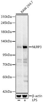
Western blot analysis of lysates from RAW 264.7 cells using NLRP3 Rabbit pAb (CAB21906) at 1:5000 dilution. Raw 264.7 cells were treated with LPS (1 μg/ml) at 37℃ for 8 hours. Secondary antibody: HRP-conjugated Goat anti-Rabbit IgG (H+L) (CABS014) at 1:10000 dilution. Lysates/proteins: 25 μg per lane. Blocking buffer: 3% nonfat dry milk in TBST. Detection: ECL Basic Kit (AbGn00020). Exposure time: 1s.
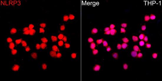
Immunofluorescence analysis of THP-1 cells using NLRP3 Rabbit pAb(CAB21906) at a dilution of 1:200 (40x lens). Secondary antibody:Cy3 Goat Anti-Rabbit IgG (H+L)(CABS007) at 1:500 dilution. Blue: DAPI for nuclear staining.
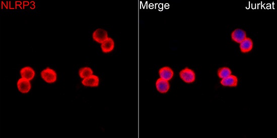
Immunofluorescence analysis of Jurkat cells using NLRP3 Rabbit pAb(CAB21906) at a dilution of 1:200 (40x lens). Secondary antibody:Cy3 Goat Anti-Rabbit IgG (H+L)(CABS007) at 1:500 dilution. Blue: DAPI for nuclear staining.
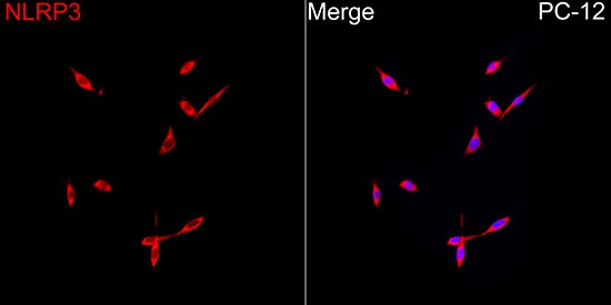
Immunofluorescence analysis of PC-12 cells using NLRP3 Rabbit pAb (CAB21906) at a dilution of 1:50 (40x lens). Secondary antibody: Cy3-conjugated Goat anti-Rabbit IgG (H+L) (CABS007) at 1:500 dilution. Blue: DAPI for nuclear staining.
 at 1:700 dilution. Raw264. 7 cells were treated by LPS (1 μg/ml) at 37℃ for 8 hours. Secondary antibody: HRP Goat Anti-Rabbit IgG (H+L) at 1:10000 dilution. Lysates/proteins: 25μg per lane. Blocking buffer: 3% nonfat dry milk in TBST.")
 at 1:700 dilution. Raw264. 7 cells were treated by LPS (1 μg/ml) at 37℃ for 8 hours. Secondary antibody: HRP Goat Anti-Rabbit IgG (H+L) at 1:10000 dilution. Lysates/proteins: 25μg per lane. Blocking buffer: 3% nonfat dry milk in TBST.")


