The NMDAR1 Monoclonal Antibody (CAB11699) is a high-quality antibody developed for reliable detection and analysis of target proteins. This antibody, produced using rabbit monoclonal technology, exhibits high specificity and sensitivity in detecting the NMDAR1 subunit in both human and animal samples.The NMDA receptor is crucial for learning, memory, and various neurological functions, making it a prime target for research into neurodegenerative disorders, psychiatric illnesses, and drug addiction. By utilizing this antibody in techniques such as Western blotting and immunofluorescence, researchers can delve deeper into the mechanisms underlying NMDA receptor function and dysfunction.
This antibody is validated for use in WB, ELISA, IF-P applications and has demonstrated reactivity against Mouse, Rat samples.
Product Name:
NMDAR1 Monoclonal Antibody
SKU:
CAB11699
Size:
20μL, 100μL
Reactivity:
Mouse, Rat
Clone Number:
ARC0684
Conjugate:
Unconjugated
Immunogen:
Synthetic peptide. This information is considered to be commercially sensitive.
The protein encoded by this gene is a critical subunit of N-methyl-D-aspartate receptors, members of the glutamate receptor channel superfamily which are heteromeric protein complexes with multiple subunits arranged to form a ligand-gated ion channel. These subunits play a key role in the plasticity of synapses, which is believed to underlie memory and learning. Cell-specific factors are thought to control expression of different isoforms, possibly contributing to the functional diversity of the subunits. Alternatively spliced transcript variants have been described.
Purification Method
Affinity purification
Gene ID
2902
RRID
AB_2861634
Buffer Information
Store at -20℃. Avoid freeze / thaw cycles. Buffer: PBS containing 50% glycerol and 0.05% BSA, preserved with proclin300 or sodium azide, pH 7.3.
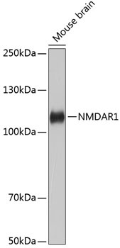
Western blot analysis of lysates from Mouse brain, using NMDAR1 Rabbit mAb (CAB11699) at 1:1000 dilution. Secondary antibody: HRP-conjugated Goat anti-Rabbit IgG (H+L) (CABS014) at 1:10000 dilution. Lysates/proteins: 25μg per lane. Blocking buffer: 3% nonfat dry milk in TBST. Detection: ECL Basic Kit (AbGn00020). Exposure time: 60s.
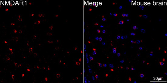
Confocal imaging of paraffin-embedded Mouse brain tissue using NMDAR1 Rabbit mAb (CAB11699, dilution 1:100) followed by a further incubation with Cy3 Goat Anti-Rabbit IgG (H+L) (CABS007, dilution 1:500) (Red). DAPI was used for nuclear staining (Blue). High pressure antigen retrieval performed with 0.01M Citrate Buffer (pH 6.0) prior to IF staining. Objective: 60x.
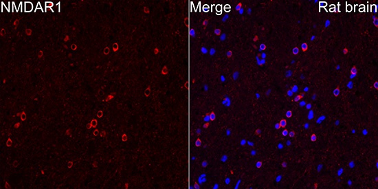
Immunofluorescence analysis of paraffin-embedded Rat brain tissue using NMDAR1 Rabbit mAb (CAB11699) at a dilution of 1:100 (40x lens). Secondary antibody:Cy3 Goat Anti-Rabbit IgG (H+L) (CABS007) at 1:500 dilution. Blue: DAPI for nuclear staining. Perform microwave antigen retrieval with 0.01 M citrate buffer (pH 6.0) prior to IF staining.