The NOB1 Antibody (CAB17690) is a high-quality antibody developed for reliable detection and analysis of target proteins. This rabbit polyclonal antibody has been validated for use in various applications, including Western blot, immunofluorescence, and immunohistochemistry, making it versatile for different experimental needs.The NOB1 antibody specifically targets the NOB1 protein, allowing for accurate detection and analysis in a variety of cell types and tissues. Its high reactivity with human samples ensures reliable results in research focused on understanding the role of NOB1 in cellular processes, such as ribosome assembly and protein degradation.
This antibody is validated for use in WB, IF/ICC, ELISA applications and has demonstrated reactivity against Human, Mouse, Rat samples.
Product Name:
NOB1 Antibody
SKU:
CAB17690
Size:
20μL, 100μL
Reactivity:
Human, Mouse, Rat
Conjugate:
Unconjugated
Immunogen:
Recombinant protein (or fragment).This information is considered to be commercially sensitive.
Recommended starting concentration is 1 μg/mL. Please optimize the concentration based on your specific assay requirements.
Synonyms:
ART-4, NOB1P, MST158, MSTP158, PSMD8BP1, NOB1
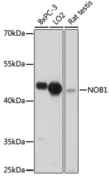
Positive Sample:
BxPC-3, LO2, rat testis
Cellular Localization:
Cytosol, Nucleoplasm.
Calculated MW:
47kDa
Observed MW:
47kDa
In yeast, over 200 protein and RNA cofactors are required for ribosome assembly, and these are generally conserved in eukaryotes. These factors orchestrate modification and cleavage of the initial 35S precursor rRNA transcript into the mature 18S, 5.8S, and 25S rRNAs, folding of the rRNA, and binding of ribosomal proteins and 5S RNA. Nob1 is involved in pre-rRNA processing. In a late cytoplasmic processing step, Nob1 cleaves a 20S rRNA intermediate at cleavage site D to produce the mature 18S rRNA (Lamanna and Karbstein, 2009 [PubMed 19706509]).
Purification Method
Affinity purification
Gene ID
28987
RRID
AB_2770608
Buffer Information
Store at -20℃. Avoid freeze / thaw cycles. Buffer: PBS with 0.01% thimerosal,50% glycerol,pH7.3.
Western blot analysis of various lysates using NOB1 Rabbit pAb (CAB17690) at 1:1000 dilution. Secondary antibody: HRP-conjugated Goat anti-Rabbit IgG (H+L) (CABS014) at 1:10000 dilution. Lysates/proteins: 25μg per lane. Blocking buffer: 3% nonfat dry milk in TBST. Detection: ECL Basic Kit (AbGn00020). Exposure time: 30s.
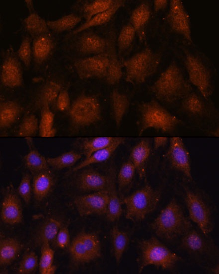
Immunofluorescence analysis of C6 cells using NOB1 Rabbit pAb (CAB17690) at dilution of 1:100. Secondary antibody: Cy3-conjugated Goat anti-Rabbit IgG (H+L) (CABS007) at 1:500 dilution. Blue: DAPI for nuclear staining.
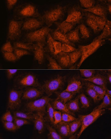
Immunofluorescence analysis of U-2 OS cells using NOB1 Rabbit pAb (CAB17690) at dilution of 1:100. Secondary antibody: Cy3-conjugated Goat anti-Rabbit IgG (H+L) (CABS007) at 1:500 dilution. Blue: DAPI for nuclear staining.