The NPHP4 Antibody (CAB8934) is a high-quality antibody developed for reliable detection and analysis of target proteins. This antibody, produced in rabbits, exhibits high reactivity with human samples and has been validated for use in immunofluorescence and immunohistochemistry applications. By binding specifically to the NPHP4 protein, this antibody enables the visualization and analysis of NPHP4 expression in a variety of cell and tissue types, making it a versatile tool for investigations in nephrology and ophthalmology research.NPHP4, also known as nephrocystin-4, plays a crucial role in the structure and function of cilia, cellular structures important for cell signaling and sensory functions.
This antibody is validated for use in WB, IF/ICC, ELISA applications and has demonstrated reactivity against Human, Mouse, Rat samples.
Product Name:
NPHP4 Antibody
SKU:
CAB8934
Size:
20μL, 100μL
Reactivity:
Human, Mouse, Rat
Conjugate:
Unconjugated
Immunogen:
Recombinant protein (or fragment).This information is considered to be commercially sensitive.
This gene encodes a protein involved in renal tubular development and function. This protein interacts with nephrocystin, and belongs to a multifunctional complex that is localized to actin- and microtubule-based structures. Mutations in this gene are associated with nephronophthisis type 4, a renal disease, and with Senior-Loken syndrome type 4, a combination of nephronophthisis and retinitis pigmentosa. Alternative splicing results in multiple transcript variants.
Purification Method
Affinity purification
Gene ID
261734
RRID
AB_2770632
Buffer Information
Store at -20℃. Avoid freeze / thaw cycles. Buffer: PBS with 0.01% thimerosal,50% glycerol,pH7.3.
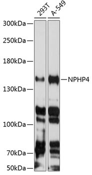
Western blot analysis of various lysates using NPHP4 Rabbit pAb (CAB8934) at 1:3000 dilution. Secondary antibody: HRP-conjugated Goat anti-Rabbit IgG (H+L) (CABS014) at 1:10000 dilution. Lysates/proteins: 25μg per lane. Blocking buffer: 3% nonfat dry milk in TBST. Detection: ECL Enhanced Kit (AbGn00021). Exposure time: 30s.
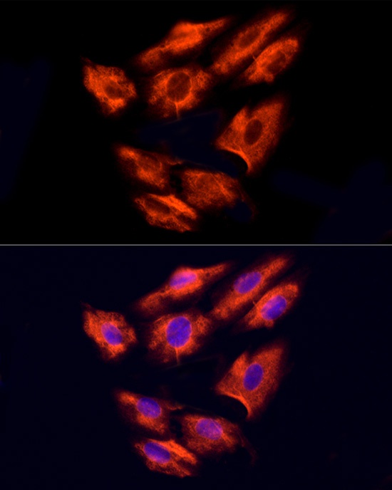
Immunofluorescence analysis of H9C2 cells using NPHP4 Rabbit pAb (CAB8934) at dilution of 100 (40x lens). Secondary antibody: Cy3-conjugated Goat anti-Rabbit IgG (H+L) (CABS007) at 1:500 dilution. Blue: DAPI for nuclear staining.
Immunofluorescence analysis of U2OS cells using NPHP4 Rabbit pAb (CAB8934) at dilution of 100 (40x lens). Secondary antibody: Cy3-conjugated Goat anti-Rabbit IgG (H+L) (CABS007) at 1:500 dilution. Blue: DAPI for nuclear staining.