The NSUN5 Antibody (CAB5992) is a high-quality antibody developed for reliable detection and analysis of target proteins. This rabbit-derived antibody is highly specific to human NSUN5 and has been validated for use in applications such as Western blotting. By binding to the NSUN5 protein, this antibody enables researchers to detect and analyze NSUN5 expression in a variety of cell types, making it an ideal choice for studies in molecular biology and RNA biology.NSUN5 is known to play a key role in regulating RNA modification processes, which are essential for ensuring proper gene expression and cellular function. Dysregulation of NSUN5 has been linked to various diseases, including cancer, neurological disorders, and developmental abnormalities.
This antibody is validated for use in WB, IHC-P, ELISA applications and has demonstrated reactivity against Human, Mouse, Rat samples.
Product Name:
NSUN5 Antibody
SKU:
CAB5992
Size:
20μL, 100μL
Reactivity:
Human, Mouse, Rat
Conjugate:
Unconjugated
Immunogen:
Recombinant protein (or fragment).This information is considered to be commercially sensitive.
This gene encodes a member of an evolutionarily conserved family of proteins that may function as methyltransferases. This gene is located in a larger region of chromosome 7 that is deleted in Williams-Beuren syndrome, a multisystem developmental disorder. There are two pseudogenes for this gene located in the same region of chromosome 7. Alternative splicing results in multiple transcript variants encoding different isoforms.
Purification Method
Affinity purification
Gene ID
55695
RRID
AB_2770675
Buffer Information
Store at -20℃. Avoid freeze / thaw cycles. Buffer: PBS containing 50% glycerol, preserved with proclin300 or sodium azide, pH 7.3.
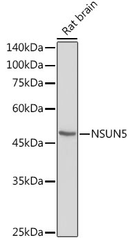
Western blot analysis of lysates from Rat brain, using NSUN5 Rabbit pAb (CAB5992) at 1:1000 dilution. Secondary antibody: HRP-conjugated Goat anti-Rabbit IgG (H+L) (CABS014) at 1:10000 dilution. Lysates/proteins: 25μg per lane. Blocking buffer: 3% nonfat dry milk in TBST. Detection: ECL Basic Kit (AbGn00020). Exposure time: 90s.
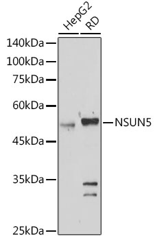
Western blot analysis of various lysates using NSUN5 Rabbit pAb (CAB5992) at 1:1000 dilution. Secondary antibody: HRP-conjugated Goat anti-Rabbit IgG (H+L) (CABS014) at 1:10000 dilution. Lysates/proteins: 25μg per lane. Blocking buffer: 3% nonfat dry milk in TBST. Detection: ECL Basic Kit (AbGn00020). Exposure time: 180s.
Immunohistochemistry analysis of paraffin-embedded Mouse spinal cord using NSUN5 Rabbit pAb ( CAB5992) at dilution of 1:100 (40x lens). High pressure antigen retrieval performed with 0.01M Citrate buffer (pH 6.0) prior to IHC staining.
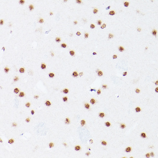
Immunohistochemistry analysis of paraffin-embedded Rat brain using NSUN5 Rabbit pAb ( CAB5992) at dilution of 1:100 (40x lens). High pressure antigen retrieval performed with 0.01M Citrate buffer (pH 6.0) prior to IHC staining.