The NTS Antibody (CAB12012) is a high-quality antibody developed for reliable detection and analysis of target proteins. This antibody, generated in rabbits, exhibits high reactivity with human samples and has been validated for use in Western blot applications.The NTS Polyclonal Antibody enables specific binding to the NTS protein, allowing for precise detection and analysis in a variety of cell types.
This antibody is validated for use in WB, IHC-P, ELISA applications and has demonstrated reactivity against Human, Mouse, Rat samples.
Product Name:
NTS Antibody
SKU:
CAB12012
Size:
20μL, 100μL
Reactivity:
Human, Mouse, Rat
Conjugate:
Unconjugated
Immunogen:
Synthetic peptide. This information is considered to be commercially sensitive.
Recommended starting concentration is 1 μg/mL. Please optimize the concentration based on your specific assay requirements.
Synonyms:
NN, NT, NT/N, NTS1, NMN-125, NTS
Positive Sample:
293T
Cellular Localization:
Cytoplasmic Vesicle, Secreted, Secretory Vesicle.
Calculated MW:
20kDa
Observed MW:
20kDa
This gene encodes a common precursor for two peptides, neuromedin N and neurotensin. Neurotensin is a secreted tridecapeptide, which is widely distributed throughout the central nervous system, and may function as a neurotransmitter or a neuromodulator. It may be involved in dopamine-associated pathophysiological events, in the maintenance of gut structure and function, and in the regulation of fat metabolism. Neurotensin also exhibits antimicrobial activity against bacteria and fungi. Tissue-specific processing may lead to the formation in some tissues of larger forms of neuromedin N and neurotensin. The large forms may represent more stable peptides that are also biologically active.
Purification Method
Affinity purification
Gene ID
4922
RRID
AB_2758928
Buffer Information
Store at -20℃. Avoid freeze / thaw cycles. Buffer: PBS containing 50% glycerol, preserved with proclin300 or sodium azide, pH 7.3.
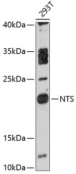
Western blot analysis of lysates from 293T cells, using NTS Rabbit pAb (CAB12012) at 1:3000 dilution. Secondary antibody: HRP-conjugated Goat anti-Rabbit IgG (H+L) (CABS014) at 1:10000 dilution. Lysates/proteins: 25μg per lane. Blocking buffer: 3% nonfat dry milk in TBST. Detection: ECL Basic Kit (AbGn00020). Exposure time: 90s.
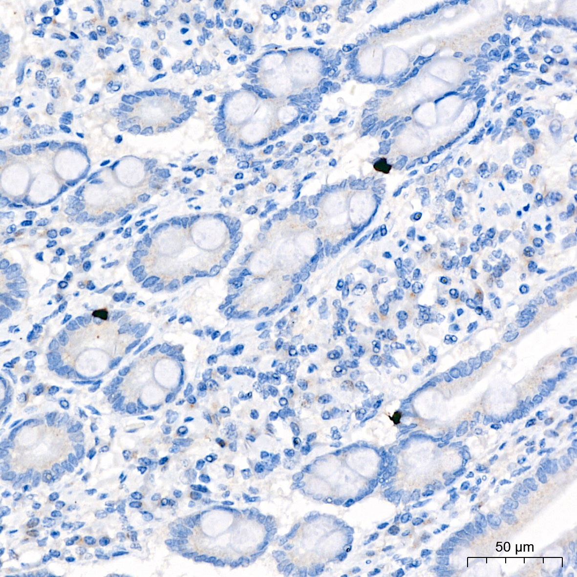
Immunohistochemistry analysis of paraffin-embedded Human small intestine tissue using NTS Rabbit pAb (CAB12012) at a dilution of 1:500 (40x lens). High pressure antigen retrieval performed with 0.01M Citrate Buffer (pH 6.0) prior to IHC staining.