The NUAK1 Antibody (CAB17333) is a high-quality antibody developed for reliable detection and analysis of target proteins. Enables p53 binding activity and protein serine/threonine kinase activity. Involved in several processes, including protein phosphorylation; regulation of cellular senescence; and regulation of myosin-light-chain-phosphatase activity. Located in cytoplasm; microtubule cytoskeleton; and nuclear lumen.
This antibody is validated for use in WB, ELISA applications and has demonstrated reactivity against Human, Rat samples.
Product Name:
NUAK1 Antibody
SKU:
CAB17333
Size:
100μL, 20μL
Reactivity:
Human, Rat
Conjugate:
Unconjugated
Immunogen:
Recombinant protein (or fragment).This information is considered to be commercially sensitive.
Tested Applications:
WBELISA
Recommended Dilution:
WB
1:200 - 1:2000
ELISA
Recommended starting concentration is 1 μg/mL. Please optimize the concentration based on your specific assay requirements.
Synonyms:
ARK5, NUAK1
Positive Sample:
rat heart
Cellular Localization:
Cytoplasm, Nucleus.
Calculated MW:
74kDa
Observed MW:
74kDa
Enables p53 binding activity and protein serine/threonine kinase activity. Involved in several processes, including protein phosphorylation; regulation of cellular senescence; and regulation of myosin-light-chain-phosphatase activity. Located in cytoplasm; microtubule cytoskeleton; and nuclear lumen.
Purification Method
Affinity purification
Gene ID
9891
RRID
AB_2770680
Buffer Information
Store at -20℃. Avoid freeze / thaw cycles. Buffer: PBS with 0.01% thimerosal,50% glycerol,pH7.3.
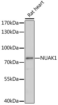
Western blot analysis of lysates from rat heart, using NUAK1 Rabbit pAb (CAB17333) at 1:1000 dilution. Secondary antibody: HRP-conjugated Goat anti-Rabbit IgG (H+L) (AS014) at 1:10000 dilution. Lysates/proteins: 25μg per lane. Blocking buffer: 3% nonfat dry milk in TBST. Detection: ECL Basic Kit (AbGn00020). Exposure time: 30s.