The NUMA1 Monoclonal Antibody (CAB4416) is a high-quality antibody developed for reliable detection and analysis of target proteins. This antibody, generated in rabbits, exhibits high specificity and sensitivity when used with human samples, making it an ideal choice for immunofluorescence, immunoprecipitation, and immunohistochemistry applications.NUMA, or Nuclear Mitotic Apparatus protein, plays a crucial role in organizing microtubules during mitosis and ensuring accurate chromosome segregation. Dysregulation of NUMA has been linked to various diseases, including cancer and developmental disorders.
This antibody is validated for use in WB, IHC-P, ELISA applications and has demonstrated reactivity against Human, Mouse, Rat samples.
Product Name:
NUMA1 Monoclonal Antibody
SKU:
CAB4416
Size:
20μL, 100μL
Reactivity:
Human, Mouse, Rat
Clone Number:
ARC1000
Conjugate:
Unconjugated
Immunogen:
Synthetic peptide. This information is considered to be commercially sensitive.
Sequence:
TPES KKAT SCFP RPMT PRDR HEGR KQST TEAQ KKAA PAST KQAD RRQS MAFS ILNT PKKL GNSL LRRG ASKK ALSK ASPN TRSG TRRS PRIA TTTA SAAT A
Tested Applications:
WBIHC-PELISA
Recommended Dilution:
WB
1:500 - 1:2000
IHC-P
1:50 - 1:200
ELISA
Recommended starting concentration is 1 μg/mL. Please optimize the concentration based on your specific assay requirements.
Synonyms:
NUMA, NMP-22, NUMA1
Positive Sample:
A-549, Mouse lung
Cellular Localization:
Chromosome, Cytoplasm, Nucleus Matrix, Cytoskeleton, Spindle Pole.
Calculated MW:
238kDa
Observed MW:
238kDa
This gene encodes a large protein that forms a structural component of the nuclear matrix. The encoded protein interacts with microtubules and plays a role in the formation and organization of the mitotic spindle during cell division. Chromosomal translocation of this gene with the RARA (retinoic acid receptor, alpha) gene on chromosome 17 have been detected in patients with acute promyelocytic leukemia. Alternative splicing results in multiple transcript variants.
Purification Method
Affinity purification
Gene ID
4926
RRID
AB_2863270
Buffer Information
Store at -20℃. Avoid freeze / thaw cycles. Buffer: PBS containing 50% glycerol and 0.05% BSA, preserved with proclin300 or sodium azide, pH 7.3.
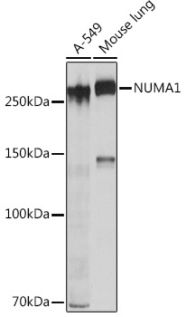
Western blot analysis of various lysates using NUMA1 Rabbit mAb (CAB4416) at 1:1000 dilution. Secondary antibody: HRP-conjugated Goat anti-Rabbit IgG (H+L) (CABS014) at 1:10000 dilution. Lysates/proteins: 25 μg per lane. Blocking buffer: 3% nonfat dry milk in TBST. Detection: ECL Basic Kit (AbGn00020). Exposure time: 10s.
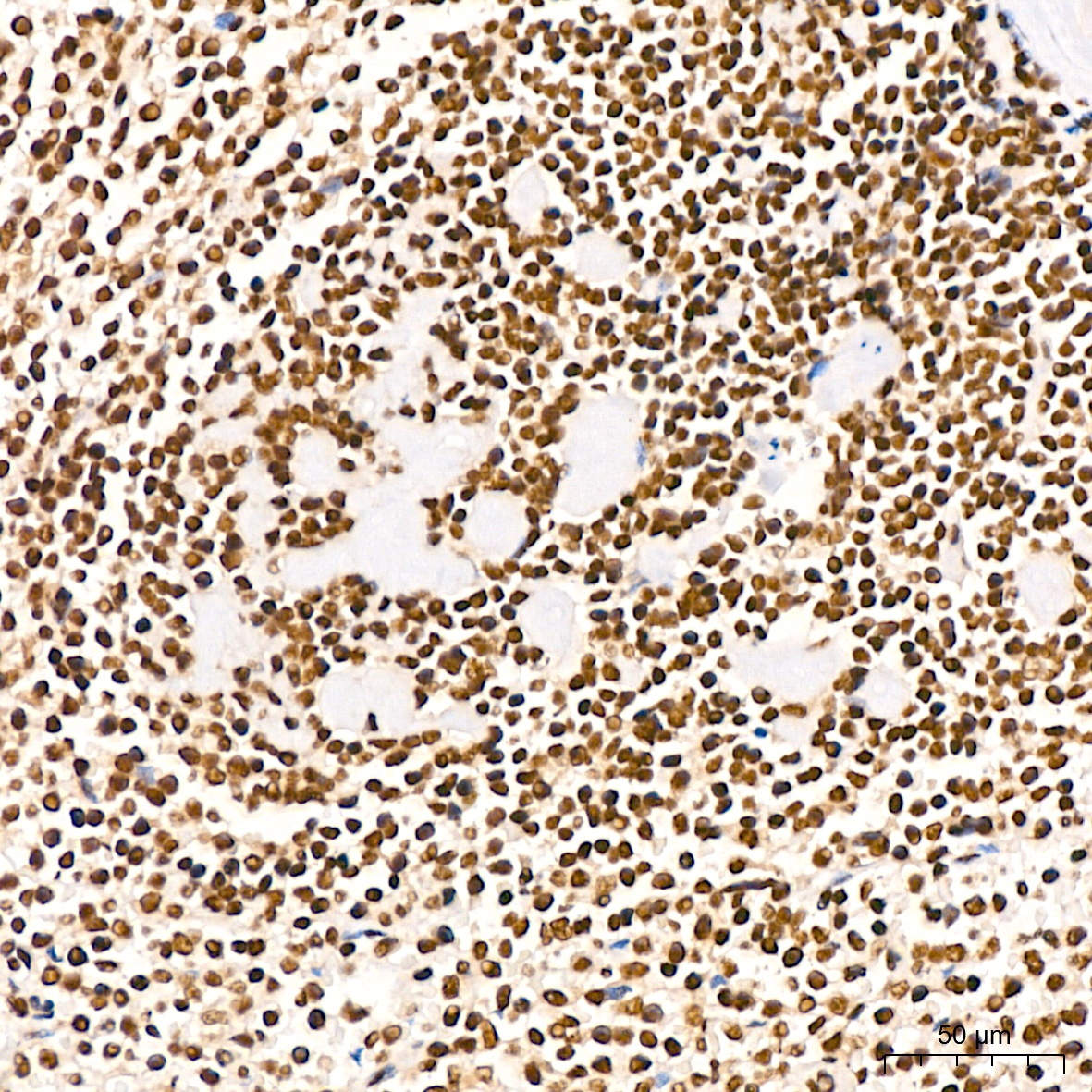
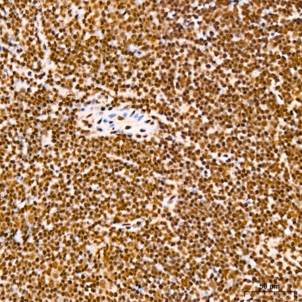
Immunohistochemistry analysis of paraffin-embedded Human spleen tissue using NUMA1 Rabbit mAb (CAB4416) at a dilution of 1:200 (40x lens). High pressure antigen retrieval performed with 0.01M Citrate buffer (pH 6.0) prior to IHC staining.
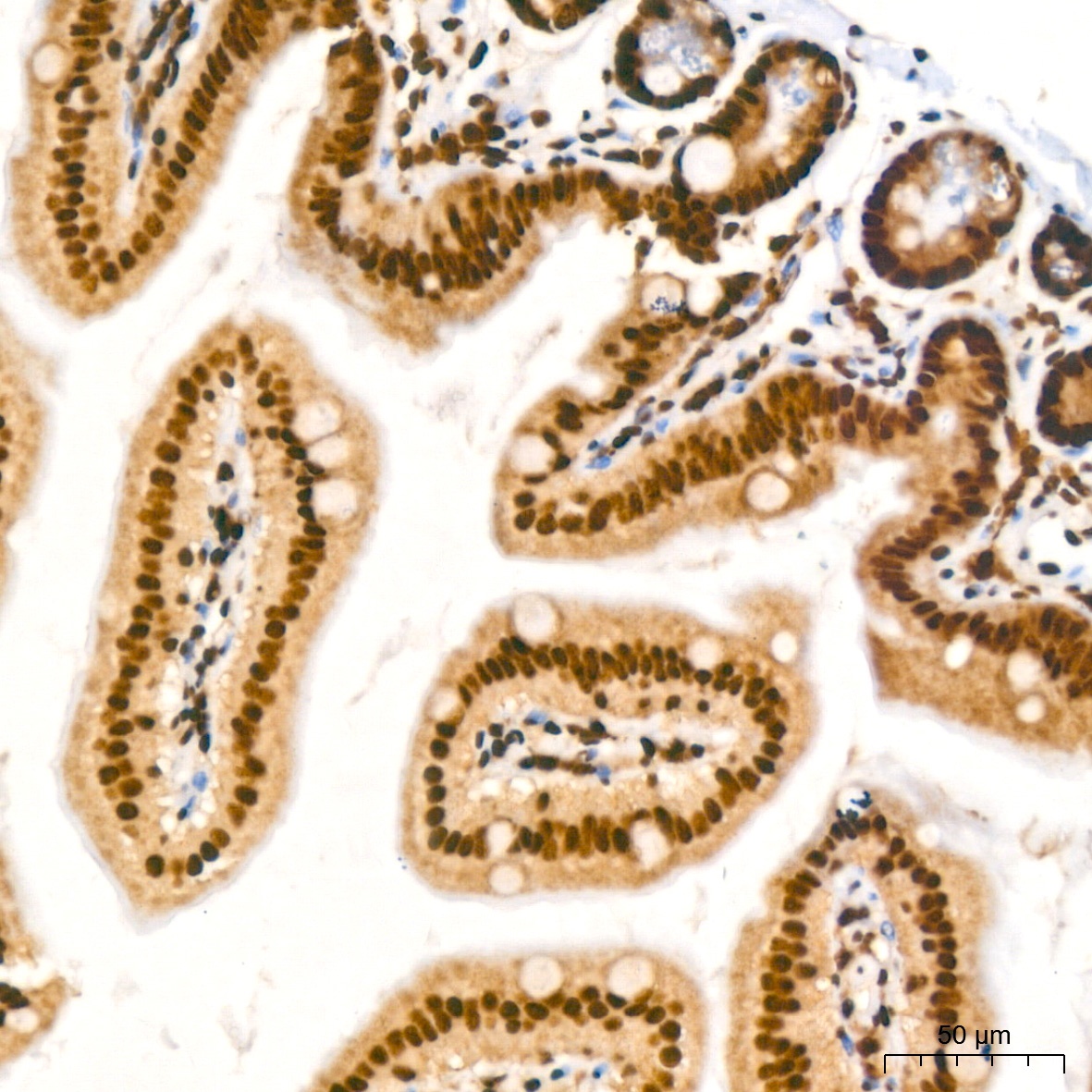
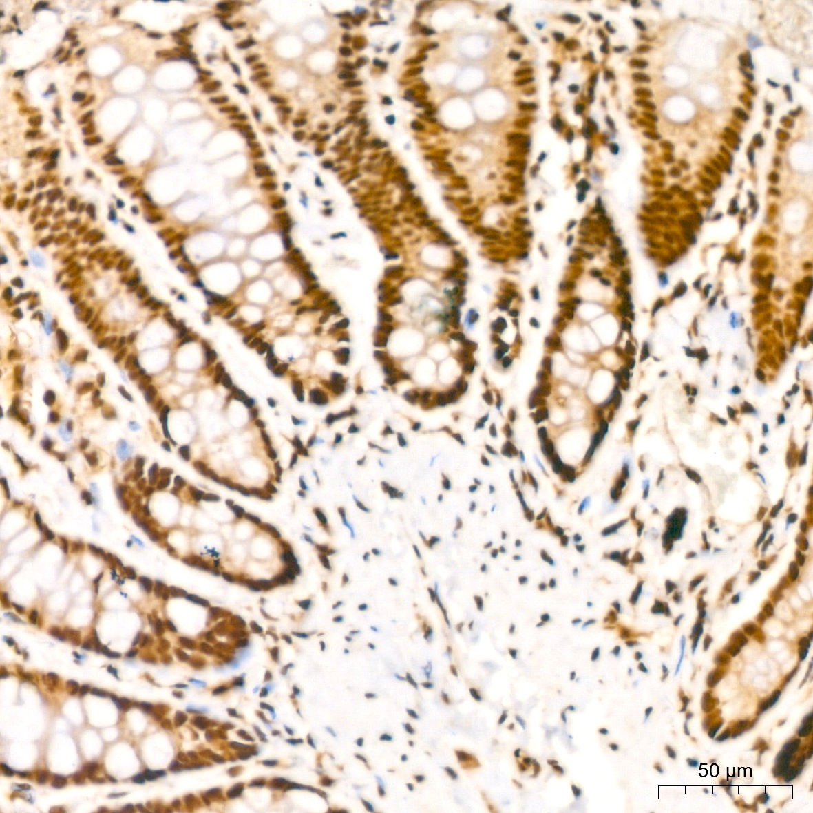
Immunohistochemistry analysis of paraffin-embedded Mouse intestin tissue using NUMA1 Rabbit mAb (CAB4416) at a dilution of 1:200 (40x lens). High pressure antigen retrieval performed with 0.01M Citrate buffer (pH 6.0) prior to IHC staining.
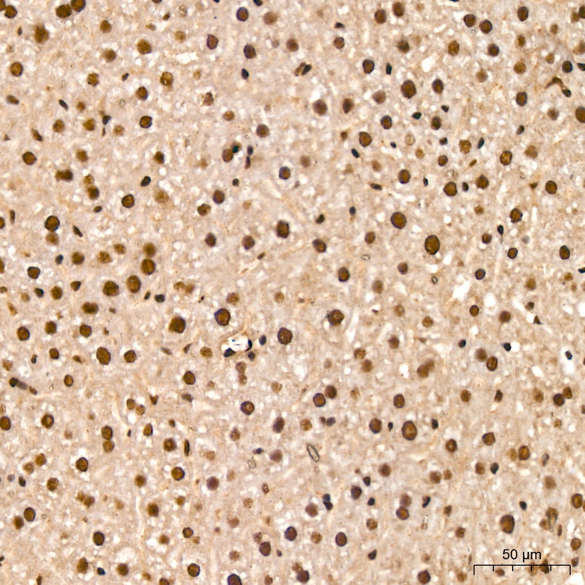
Immunohistochemistry analysis of paraffin-embedded Mouse liver tissue using NUMA1 Rabbit mAb (CAB4416) at a dilution of 1:200 (40x lens). High pressure antigen retrieval performed with 0.01M Citrate buffer (pH 6.0) prior to IHC staining.
Immunohistochemistry analysis of paraffin-embedded Mouse spleen tissue using NUMA1 Rabbit mAb (CAB4416) at a dilution of 1:200 (40x lens). High pressure antigen retrieval performed with 0.01M Citrate buffer (pH 6.0) prior to IHC staining.
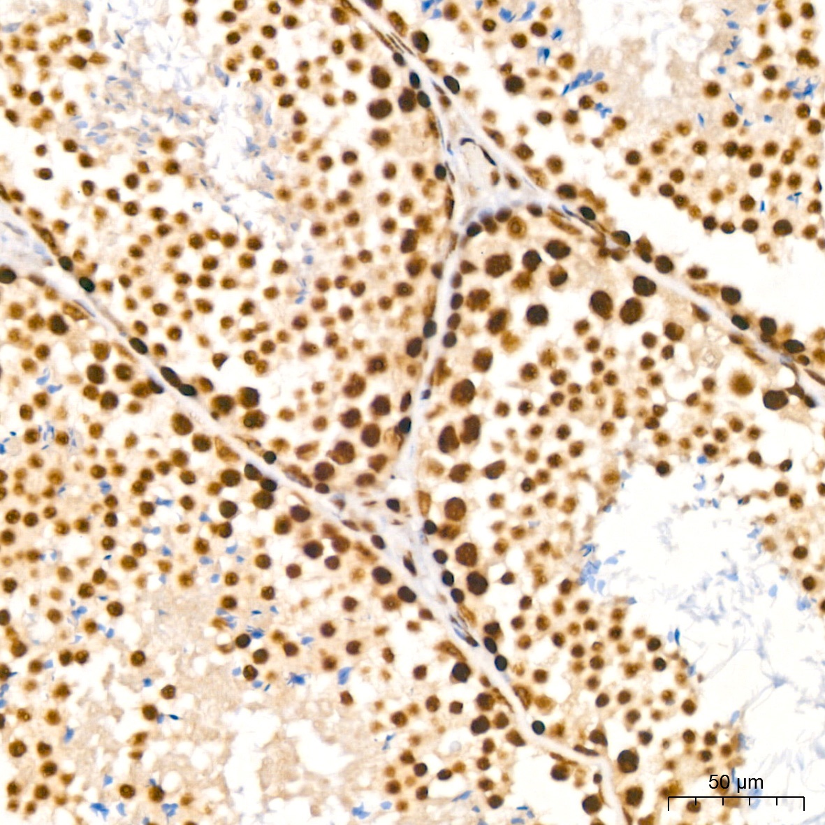
Immunohistochemistry analysis of paraffin-embedded Mouse testis tissue using NUMA1 Rabbit mAb (CAB4416) at a dilution of 1:200 (40x lens). High pressure antigen retrieval performed with 0.01M Citrate buffer (pH 6.0) prior to IHC staining.
Immunohistochemistry analysis of paraffin-embedded Rat colon tissue using NUMA1 Rabbit mAb (CAB4416) at a dilution of 1:200 (40x lens). High pressure antigen retrieval performed with 0.01M Citrate buffer (pH 6.0) prior to IHC staining.
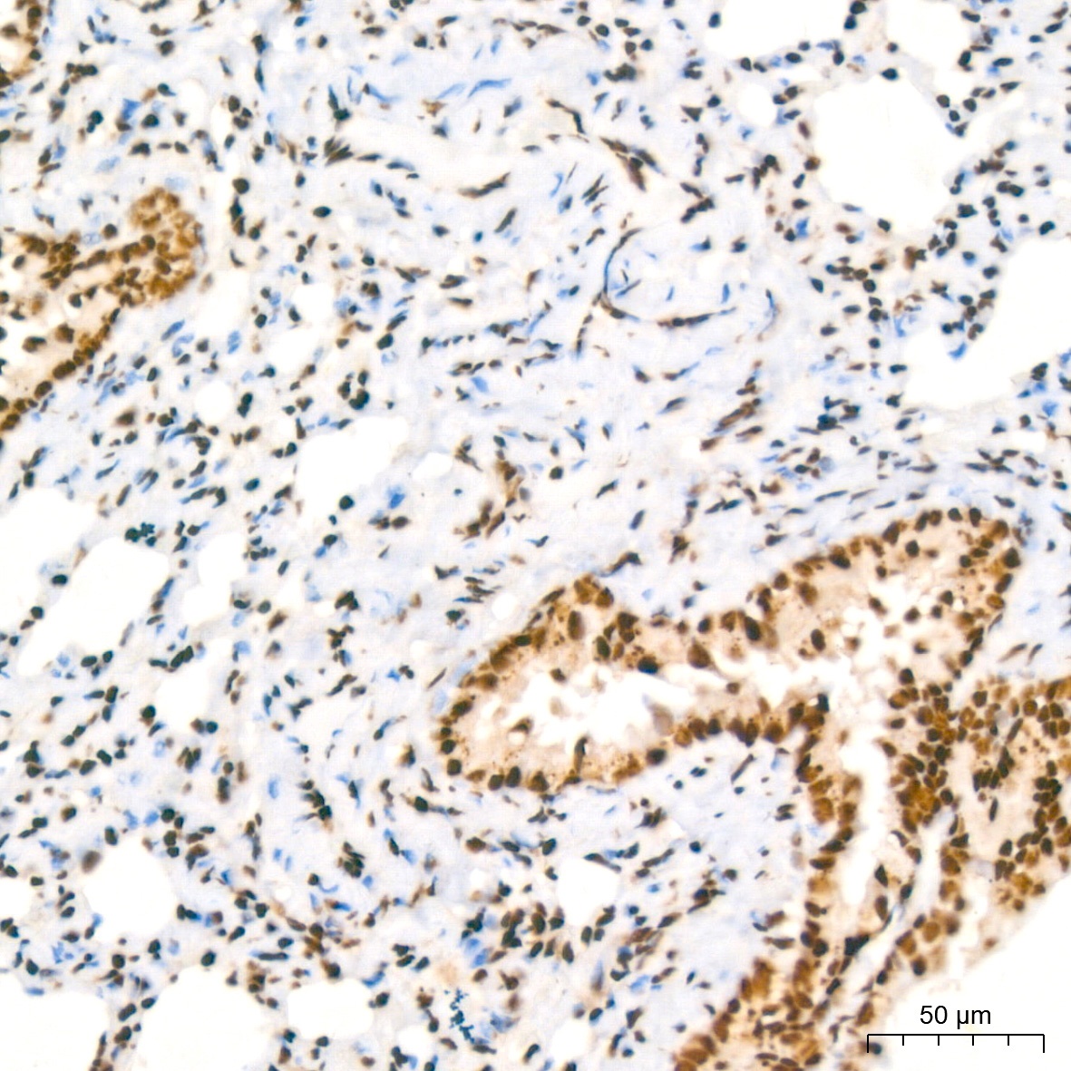
Immunohistochemistry analysis of paraffin-embedded Rat lung tissue using NUMA1 Rabbit mAb (CAB4416) at a dilution of 1:200 (40x lens). High pressure antigen retrieval performed with 0.01M Citrate buffer (pH 6.0) prior to IHC staining.