The ODC1 Antibody (CAB1948) is a high-quality antibody developed for reliable detection and analysis of target proteins. This antibody, generated in rabbits, demonstrates high reactivity with human samples and has been validated for use in Western blot applications.ODC1, also known as ornithine decarboxylase, plays a critical role in cell growth and proliferation by catalyzing the decarboxylation of ornithine to form putrescine. Dysregulation of ODC1 has been linked to various diseases, including cancer and neurodegenerative disorders.
This antibody is validated for use in WB, IF/ICC, ELISA applications and has demonstrated reactivity against Human, Mouse, Rat samples.
Product Name:
ODC1 Antibody
SKU:
CAB1948
Size:
20μL, 100μL
Reactivity:
Human, Mouse, Rat
Conjugate:
Unconjugated
Immunogen:
Recombinant protein (or fragment).This information is considered to be commercially sensitive.
Recommended starting concentration is 1 μg/mL. Please optimize the concentration based on your specific assay requirements.
Synonyms:
ODC, BABS, NEDBA, NEDBIA, ODC1
Positive Sample:
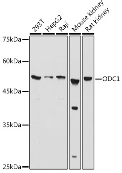
293T, HepG2, Raji, Mouse kidney, Rat kidney
Cellular Localization:
Cytoplasm, Cytosol.
Calculated MW:
51kDa
Observed MW:
51kDa
This gene encodes the rate-limiting enzyme of the polyamine biosynthesis pathway which catalyzes ornithine to putrescine. The activity level for the enzyme varies in response to growth-promoting stimuli and exhibits a high turnover rate in comparison to other mammalian proteins. Originally localized to both chromosomes 2 and 7, the gene encoding this enzyme has been determined to be located on 2p25, with a pseudogene located on 7q31-qter. Multiple alternatively spliced transcript variants encoding distinct isoforms have been identified.
Purification Method
Affinity purification
Gene ID
4953
RRID
AB_2763974
Buffer Information
Store at -20℃. Avoid freeze / thaw cycles. Buffer: PBS containing 50% glycerol, preserved with proclin300 or sodium azide, pH 7.3.
Western blot analysis of various lysates using ODC1 Rabbit pAb (CAB1948) at 1:1000 dilution. Secondary antibody: HRP-conjugated Goat anti-Rabbit IgG (H+L) (CABS014) at 1:10000 dilution. Lysates/proteins: 25μg per lane. Blocking buffer: 3% nonfat dry milk in TBST. Detection: ECL Basic Kit (AbGn00020). Exposure time: 10s.
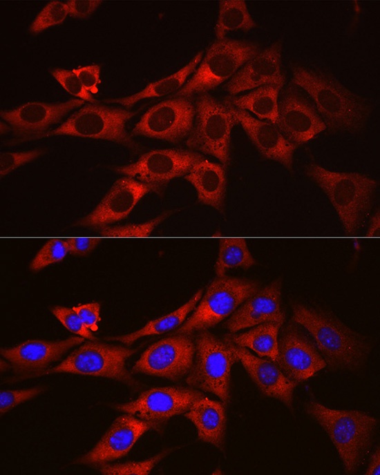
Immunofluorescence analysis of NIH/3T3 cells using ODC1 Rabbit pAb (CAB1948) at dilution of 1:100 (40x lens). Secondary antibody: Cy3-conjugated Goat anti-Rabbit IgG (H+L) (CABS007) at 1:500 dilution. Blue: DAPI for nuclear staining.
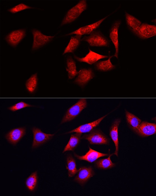
Immunofluorescence analysis of U2OS cells using ODC1 Rabbit pAb (CAB1948) at dilution of 1:50 (40x lens). Secondary antibody: Cy3-conjugated Goat anti-Rabbit IgG (H+L) (CABS007) at 1:500 dilution. Blue: DAPI for nuclear staining.