The OXSR1 Antibody (CAB15126) is a high-quality antibody developed for reliable detection and analysis of target proteins. This antibody, produced in rabbits, is highly specific for OXSR1 and has been validated for use in various research applications, including Western blot and immunohistochemistry.OXSR1 plays a critical role in regulating cell growth, proliferation, and survival, making it a key target in studies related to cancer, neurological disorders, and cardiovascular diseases.
This antibody is validated for use in WB, IF/ICC, ELISA applications and has demonstrated reactivity against Human, Mouse, Rat samples.
Product Name:
OXSR1 Antibody
SKU:
CAB15126
Size:
20μL, 100μL
Reactivity:
Human, Mouse, Rat
Conjugate:
Unconjugated
Immunogen:
Synthetic peptide. This information is considered to be commercially sensitive.
Recommended starting concentration is 1 μg/mL. Please optimize the concentration based on your specific assay requirements.
Synonyms:
OSR1, OXSR1
Positive Sample:
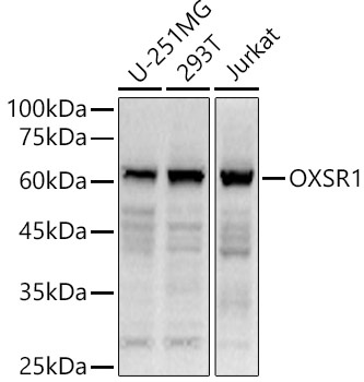
U-251MG, 293T, Jurkat
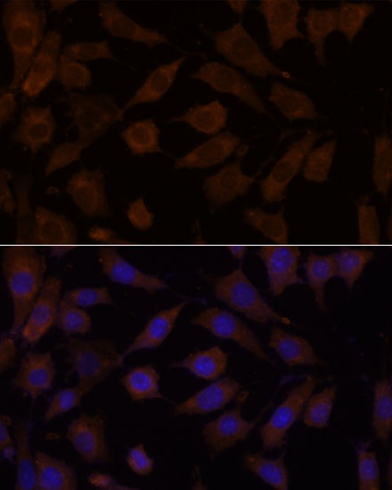
Cellular Localization:
Cytoplasm.
Calculated MW:
58kDa
Observed MW:
60kDa
The product of this gene belongs to the Ser/Thr protein kinase family of proteins. It regulates downstream kinases in response to environmental stress, and may play a role in regulating the actin cytoskeleton.
Purification Method
Affinity purification
Gene ID
9943
RRID
AB_2762011
Buffer Information
Store at -20℃. Avoid freeze / thaw cycles. Buffer: PBS with 0.01% thimerosal,50% glycerol,pH7.3.
Western blot analysis of various lysates, using OXSR1 Rabbit pAb (CAB15126) at 1:1000 dilution. Secondary antibody: HRP-conjugated Goat anti-Rabbit IgG (H+L) (CABS014) at 1:10000 dilution. Lysates/proteins: 25μg per lane. Blocking buffer: 3% nonfat dry milk in TBST. Detection: ECL Basic Kit (AbGn00020). Exposure time: 10s.
Immunofluorescence analysis of L929 cells using OXSR1 Rabbit pAb (CAB15126) at dilution of 1:100. Secondary antibody: Cy3-conjugated Goat anti-Rabbit IgG (H+L) (CABS007) at 1:500 dilution. Blue: DAPI for nuclear staining.