The P2RX4 Antibody (CAB6682) is a high-quality antibody developed for reliable detection and analysis of target proteins. This antibody, generated in rabbits, exhibits high reactivity with human samples and is validated for use in Western blot applications. By binding to the P2X4 receptor protein, this antibody enables the detection and analysis of P2X4 in various cell types, making it an ideal choice for studies in neuroscience, pharmacology, and immunology.P2X4 is a member of the P2X receptor family, which are ligand-gated ion channels activated by extracellular ATP.
This antibody is validated for use in WB, IF/ICC, ELISA applications and has demonstrated reactivity against Human, Mouse samples.
Product Name:
P2RX4 Antibody
SKU:
CAB6682
Size:
20μL, 100μL
Reactivity:
Human, Mouse
Conjugate:
Unconjugated
Immunogen:
Recombinant protein (or fragment).This information is considered to be commercially sensitive.
Recommended starting concentration is 1 μg/mL. Please optimize the concentration based on your specific assay requirements.
Synonyms:
P2X4, P2X4R, P2RX4
Positive Sample:
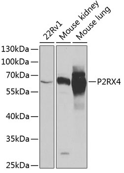
22Rv1, Mouse kidney, Mouse lung
Cellular Localization:
Membrane, Multi-Pass Membrane Protein.
Calculated MW:
43kDa
Observed MW:
62kDa
The product of this gene belongs to the family of purinoceptors for ATP. This receptor functions as a ligand-gated ion channel with high calcium permeability. The main pharmacological distinction between the members of the purinoceptor family is the relative sensitivity to the antagonists suramin and PPADS. The product of this gene has the lowest sensitivity for these antagonists. Multiple alternatively spliced transcript variants, some protein-coding and some not protein-coding, have been found for this gene.
Purification Method
Affinity purification
Gene ID
5025
RRID
AB_2767266
Buffer Information
Store at -20℃. Avoid freeze / thaw cycles. Buffer: PBS containing 50% glycerol, preserved with proclin300 or sodium azide, pH 7.3.
Western blot analysis of various lysates using P2RX4 Rabbit pAb (CAB6682) at 1:1000 dilution. Secondary antibody: HRP-conjugated Goat anti-Rabbit IgG (H+L) (CABS014) at 1:10000 dilution. Lysates/proteins: 25μg per lane. Blocking buffer: 3% nonfat dry milk in TBST. Detection: ECL Basic Kit (AbGn00020). Exposure time: 30s.
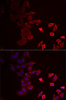
Immunofluorescence analysis of MCF7 cells using P2RX4 Rabbit pAb (CAB6682). Secondary antibody: Cy3-conjugated Goat anti-Rabbit IgG (H+L) (CABS007) at 1:500 dilution. Blue: DAPI for nuclear staining.