The pAbPC1 Antibody (CAB14872) is a high-quality antibody developed for reliable detection and analysis of target proteins. This antibody, produced using rabbits, is highly specific for human samples and has been validated for use in Western blotting applications.PABPC1, also known as poly(A)-binding protein cytoplasmic 1, plays a crucial role in mRNA stability and translation efficiency. Dysregulation of PABPC1 has been linked to various diseases, including cancer, neurodegenerative disorders, and viral infections.
This antibody is validated for use in WB, IHC-P, IF/ICC, ELISA applications and has demonstrated reactivity against Human, Mouse, Rat samples.
Product Name:
pAbPC1 Antibody
SKU:
CAB14872
Size:
20μL, 100μL
Reactivity:
Human, Mouse, Rat
Conjugate:
Unconjugated
Immunogen:
Synthetic peptide. This information is considered to be commercially sensitive.
Recommended starting concentration is 1 μg/mL. Please optimize the concentration based on your specific assay requirements.
Synonyms:
PAB1, PABP, PABP1, PABPC2, PABPL1, pAbPC1
Positive Sample:
NIH/3T3, PC-12
Cellular Localization:
Cytoplasm, Nucleus.
Calculated MW:
71kDa
Observed MW:
75kDa
This gene encodes a poly(A) binding protein. The protein shuttles between the nucleus and cytoplasm and binds to the 3' poly(A) tail of eukaryotic messenger RNAs via RNA-recognition motifs. The binding of this protein to poly(A) promotes ribosome recruitment and translation initiation; it is also required for poly(A) shortening which is the first step in mRNA decay. The gene is part of a small gene family including three protein-coding genes and several pseudogenes.
Purification Method
Affinity purification
Gene ID
26986
RRID
AB_2761752
Buffer Information
Store at -20℃. Avoid freeze / thaw cycles. Buffer: PBS containing 50% glycerol, preserved with proclin300 or sodium azide, pH 7.3.
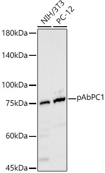
Western blot analysis of various lysates, using pAbPC1 Rabbit pAb (CAB14872) at 1:900 dilution. Secondary antibody: HRP-conjugated Goat anti-Rabbit IgG (H+L) (CABS014) at 1:10000 dilution. Lysates/proteins: 25μg per lane. Blocking buffer: 3% nonfat dry milk in TBST. Detection: ECL Basic Kit (AbGn00020). Exposure time: 30s.
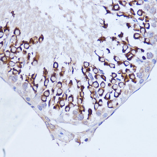
Immunohistochemistry analysis of paraffin-embedded Rat testis using pAbPC1 Rabbit pAb (CAB14872) at dilution of 1:100 (40x lens). Microwave antigen retrieval performed with 0.01M PBS Buffer (pH 7.2) prior to IHC staining.
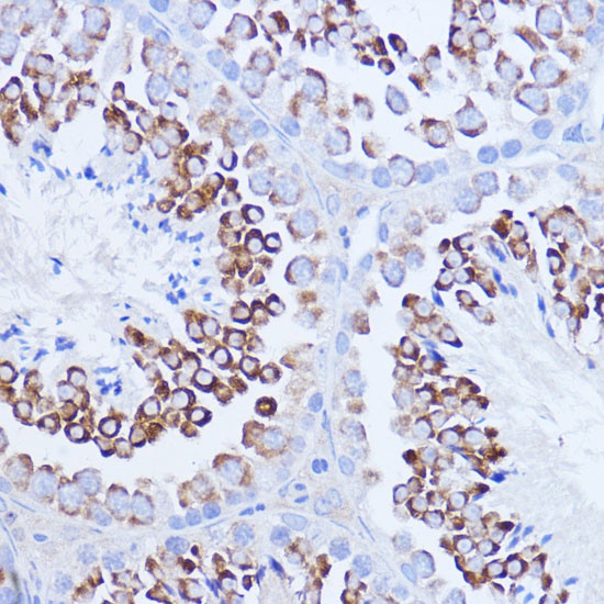
Immunohistochemistry analysis of paraffin-embedded Mouse testis using pAbPC1 Rabbit pAb (CAB14872) at dilution of 1:100 (40x lens). Microwave antigen retrieval performed with 0.01M PBS Buffer (pH 7.2) prior to IHC staining.
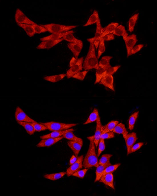
Immunofluorescence analysis of MCF7 cells using pAbPC1 Rabbit pAb (CAB14872) at dilution of 1:100 (40x lens). Secondary antibody: Cy3-conjugated Goat anti-Rabbit IgG (H+L) (CABS007) at 1:500 dilution. Blue: DAPI for nuclear staining.
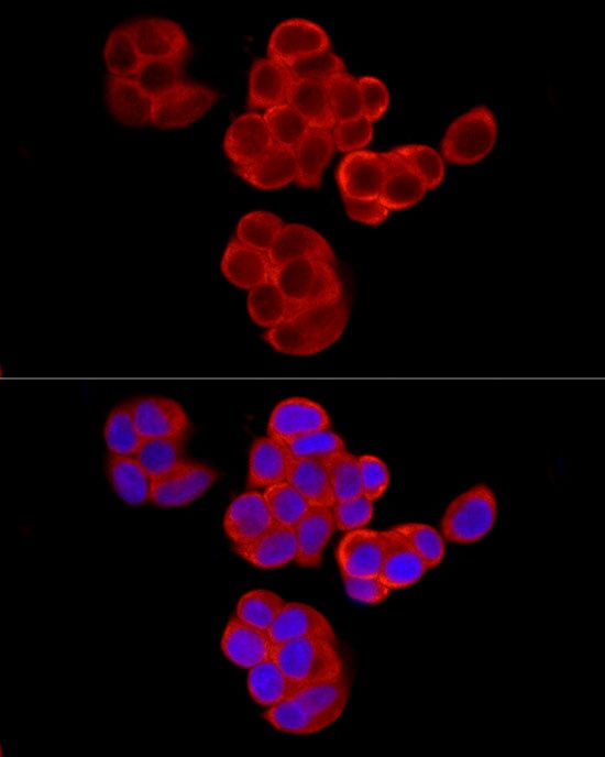
Immunofluorescence analysis of PC-12 cells using pAbPC1 Rabbit pAb (CAB14872) at dilution of 1:100 (40x lens). Secondary antibody: Cy3-conjugated Goat anti-Rabbit IgG (H+L) (CABS007) at 1:500 dilution. Blue: DAPI for nuclear staining.