The LIS1 Monoclonal Antibody (CAB3696) is a high-quality antibody developed for reliable detection and analysis of target proteins. This antibody, produced in rabbits, is highly specific to LIS1 in human samples and is validated for use in Western blotting and immunohistochemistry applications. By binding to LIS1, this antibody enables accurate detection and analysis of the protein in various biological samples, making it an ideal choice for studies in neuroscience and developmental biology.LIS1, also known as lissencephaly-1 protein, is crucial for proper brain development and neuronal function.
This antibody is validated for use in WB, IF/ICC, IP, ELISA applications and has demonstrated reactivity against Human, Mouse, Rat samples.
Product Name:
LIS1 Monoclonal Antibody
SKU:
CAB3696
Size:
20μL, 100μL
Reactivity:
Human, Mouse, Rat
Clone Number:
ARC2075
Conjugate:
Unconjugated
Immunogen:
Synthetic peptide. This information is considered to be commercially sensitive.
0.5μg-4μg antibody for 400μg-600μg extracts of whole cells
ELISA
Recommended starting concentration is 1 μg/mL. Please optimize the concentration based on your specific assay requirements.
Synonyms:
MDS, LIS1, LIS2, MDCR, NudF, PAFAH
Positive Sample:
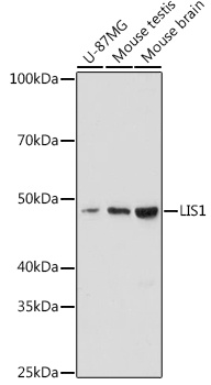
U-87MG, Mouse testis, Mouse brain
Cellular Localization:
Axon Cytoplasm, Cell Cortex, Central Region Of Growth Cone, Centrosome, Cytoplasmic Microtubule, Cytosol, Extracellular Exosome, Glutamatergic Synapse, Neuron Projection, Nuclear Envelope, Perinuclear Region Of Cytoplasm, Schaffer Collateral - Ca1 Synapse.
Calculated MW:
47kDa
Observed MW:
47kDa
This locus was identified as encoding a gene that when mutated or lost caused the lissencephaly associated with Miller-Dieker lissencephaly syndrome. This gene encodes the non-catalytic alpha subunit of the intracellular Ib isoform of platelet-activating factor acteylhydrolase, a heterotrimeric enzyme that specifically catalyzes the removal of the acetyl group at the SN-2 position of platelet-activating factor (identified as 1-O-alkyl-2-acetyl-sn-glyceryl-3-phosphorylcholine). Two other isoforms of intracellular platelet-activating factor acetylhydrolase exist: one composed of multiple subunits, the other, a single subunit. In addition, a single-subunit isoform of this enzyme is found in serum.
Purification Method
Affinity purification
Gene ID
5048
Buffer Information
Store at -20℃. Avoid freeze / thaw cycles. Buffer: PBS containing 50% glycerol and 0.05% BSA, preserved with proclin300 or sodium azide, pH 7.3.
Western blot analysis of various lysates using LIS1 Rabbit mAb (CAB3696) at 1:1000 dilution. Secondary antibody: HRP-conjugated Goat anti-Rabbit IgG (H+L) (CABS014) at 1:10000 dilution. Lysates/proteins: 25μg per lane. Blocking buffer: 3% nonfat dry milk in TBST. Detection: ECL Enhanced Kit (AbGn00021). Exposure time: 180s.
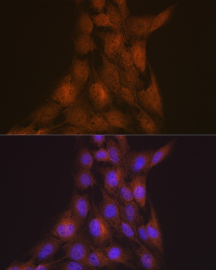
Immunofluorescence analysis of C6 cells using LIS1 Rabbit mAb (CAB3696) at dilution of 1:100 (40x lens). Secondary antibody: Cy3-conjugated Goat anti-Rabbit IgG (H+L) (CABS007) at 1:500 dilution. Blue: DAPI for nuclear staining.
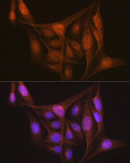
Immunofluorescence analysis of U-2 OS cells using LIS1 Rabbit mAb (CAB3696) at dilution of 1:100 (40x lens). Secondary antibody: Cy3-conjugated Goat anti-Rabbit IgG (H+L) (CABS007) at 1:500 dilution. Blue: DAPI for nuclear staining.