The Pan Cadherin Antibody (CAB18682) is a high-quality antibody developed for reliable detection and analysis of target proteins. This antibody, derived from rabbit serum, is highly specific and reactive with various species, making it suitable for a wide range of applications. It recognizes the pan-cadherin protein, a family of cell adhesion molecules essential for maintaining tissue integrity and organization.Pan-cadherin plays a crucial role in mediating cell-cell adhesion, signal transduction, and cell differentiation processes. It is involved in various physiological and pathological conditions, including embryonic development, tissue morphogenesis, and cancer metastasis.
This antibody is validated for use in WB, IP, ELISA applications and has demonstrated reactivity against Human, Mouse, Rat samples.
Product Name:
Pan Cadherin Antibody
SKU:
CAB18682
Size:
20μL, 100μL
Reactivity:
Human, Mouse, Rat
Conjugate:
Unconjugated
Immunogen:
Synthetic peptide. This information is considered to be commercially sensitive.
0.5μg-4μg antibody for 200μg-400μg extracts of whole cells
ELISA
Recommended starting concentration is 1 μg/mL. Please optimize the concentration based on your specific assay requirements.
Positive Sample:
PC-3, A-431, Mouse brain, Rat brain
Cellular Localization:
Actin Cytoskeleton, Adherens Junction, Apical Junction Complex, Cell Junction, Cytoplasm, Cytoplasmic Side Of Plasma Membrane, Endosome, Extracellular Exosome, Extracellular Region, Flotillin Complex, Glutamatergic Synapse, Lateral Plasma Membrane, Perinuclear Region Of Cytoplasm, Plasma Membrane, Trans-Golgi Network.
Observed MW:
135kDa
Cadherin is one of a class of integral-membrane glycoproteins that are involved in cell to cell attachment for preserving the integrity of all solid tissues. Cadherins have three major regions: the Ca2+ -dependent extracellular region that mediates adhesion (cadherin to cadherin) for cell to cell binding; the transmembrane region; and the cytoplasmic region that extends into the cell and interacts with catenins, which in turn are linked to the actin of the cytoskeleton. Cadherins are differentially expressed during development and in adult organs. Since many cell types express multiple cadherin subclasses simultaneously (the combination differs with cell type), it can be inferred that the adhesion properities of individual cells are thus governed by varying the combinations of cadherins. Altered expression of cadherins are involved in invasion and metastasis of tumour cells. The classical cadherins (e.g. E-, N-, and P-cadherins) are the most common family members. E-cadherin (also known as uvomorulin) is concentrated in the belt desmosome in epithelial cells; N-cadherin is found in nerve, muscle, and lens cells and helps maintain the integrity of neuronal aggregates; P-cadherin is expressed in placental and epidermal cells.
Purification Method
Affinity purification
Gene ID
999 1000 1001 1002
RRID
AB_2862418
Buffer Information
Store at -20℃. Avoid freeze / thaw cycles. Buffer: PBS with 0.01% thimerosal,50% glycerol,pH7.3.
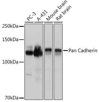
Western blot analysis of various lysates using Pan Cadherin Rabbit pAb (CAB18682) at 1:3000 dilution. Secondary antibody: HRP-conjugated Goat anti-Rabbit IgG (H+L) (CABS014) at 1:10000 dilution. Lysates/proteins: 25μg per lane. Blocking buffer: 3% nonfat dry milk in TBST. Detection: ECL Basic Kit (AbGn00020). Exposure time: 1s.
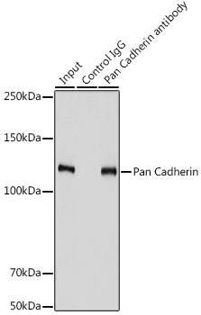
Immunoprecipitation analysis of 900 μg extracts of PC-3 cells using 3 μg Pan Cadherin antibody (CAB18682). Western blot was performed from the immunoprecipitate using Pan Cadherin antibody (CAB18682) at a dilution of 1:3000.