The Parvalbumin (PVALB) Monoclonal Antibody (CAB19098) is a high-quality antibody developed for reliable detection and analysis of target proteins. The protein encoded by this gene is a high affinity calcium ion-binding protein that is structurally and functionally similar to calmodulin and troponin C. The encoded protein is thought to be involved in muscle relaxation. Alternative splicing results in multiple transcript variants.
This antibody is validated for use in WB, IHC-P, ELISA, IF-P applications and has demonstrated reactivity against Human, Mouse, Rat samples.
Product Name:
Parvalbumin (PVALB) Monoclonal Antibody
SKU:
CAB19098
Size:
100μL, 20μL
Reactivity:
Human, Mouse, Rat
Clone Number:
ARC0385
Conjugate:
Unconjugated
Immunogen:
Recombinant protein (or fragment).This information is considered to be commercially sensitive.
Tested Applications:
WBIHC-PELISAIF-P
Recommended Dilution:
WB
1:1000 - 1:6000
IF-P
1:100 - 1:1000
IHC-P
1:200 - 1:2000
ELISA
Recommended starting concentration is 1 μg/mL. Please optimize the concentration based on your specific assay requirements.
Synonyms:
D22S749, Parvalbumin (PVALB)
Positive Sample:
Mouse skeletal muscle, Rat skeletal muscle
Cellular Localization:
Axon, Cytoplasm, Nucleus, Synapse.
Calculated MW:
12kDa
Observed MW:
12kDa
The protein encoded by this gene is a high affinity calcium ion-binding protein that is structurally and functionally similar to calmodulin and troponin C. The encoded protein is thought to be involved in muscle relaxation. Alternative splicing results in multiple transcript variants.
Purification Method
Affinity purification
Gene ID
5816
RRID
AB_2862590
Buffer Information
Store at -20℃. Avoid freeze / thaw cycles. Buffer: PBS containing 50% glycerol and 0.05% BSA, preserved with proclin300 or sodium azide, pH 7.3.
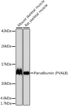
Western blot analysis of various lysates using Parvalbumin (PVALB) Rabbit mAb (CAB19098) at 1:1000 dilution. Secondary antibody: HRP-conjugated Goat anti-Rabbit IgG (H+L) (AS014) at 1:10000 dilution. Lysates/proteins: 25μg per lane. Blocking buffer: 3% nonfat dry milk in TBST. Detection: ECL Basic Kit (AbGn00020). Exposure time: 1s.
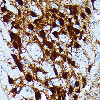
Immunohistochemistry analysis of paraffin-embedded Mouse brain tissue using Parvalbumin (PVALB) Rabbit mAb (CAB19098) at dilution of 1:200 (40x lens). Microwave antigen retrieval performed with 0.01M PBS Buffer (pH 7.2) prior to IHC staining.
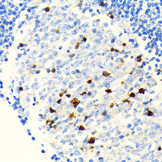
Immunohistochemistry analysis of paraffin-embedded Human appendix tissue using Parvalbumin (PVALB) Rabbit mAb (CAB19098) at dilution of 1:200 (40x lens). Microwave antigen retrieval performed with 0.01M PBS Buffer (pH 7.2) prior to IHC staining.
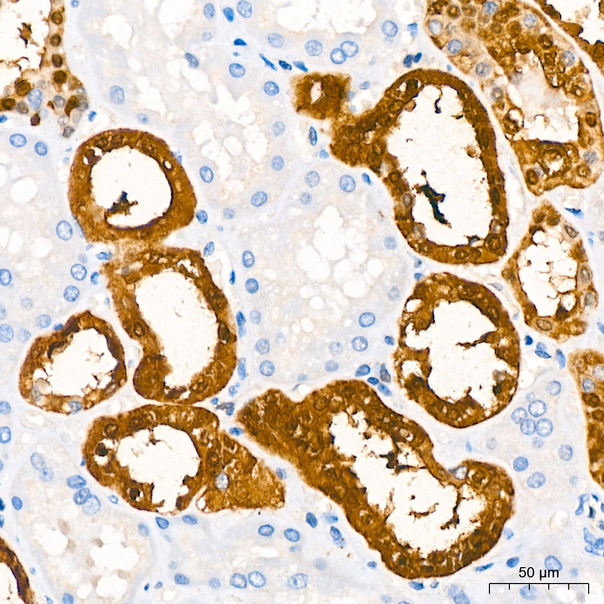
Immunohistochemistry analysis of paraffin-embedded Human kidney tissue using Parvalbumin (PVALB) Rabbit mAb (CAB19098) at dilution of 1:200 (40x lens). High pressure antigen retrieval performed with 0.01M Citrate Buffer (pH 6.0) prior to IHC staining.
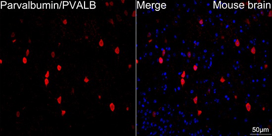
Confocal imaging of paraffin-embedded Mouse brain tissue using Parvalbumin/PVALB Rabbit mAb (CAB19098, dilution 1:100) followed by a further incubation with Cy3 Goat Anti-Rabbit IgG (H+L) (AS007, dilution 1:500) (Red). DAPI was used for nuclear staining (Blue). Microwave antigen retrieval performed with 0.01M Citrate Buffer (pH 6.0) prior to IF staining. Objective: 40x.
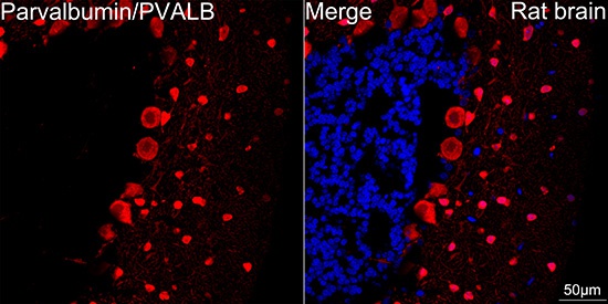
Confocal imaging of paraffin-embedded Rat brain tissue using Parvalbumin/PVALB Rabbit mAb (CAB19098, dilution 1:100) followed by a further incubation with Cy3 Goat Anti-Rabbit IgG (H+L) (AS007, dilution 1:500) (Red). DAPI was used for nuclear staining (Blue). Microwave antigen retrieval performed with 0.01M Citrate Buffer (pH 6.0) prior to IF staining. Objective: 40x.