The PAX1 Antibody (CAB17513) is a high-quality antibody developed for reliable detection and analysis of target proteins. This antibody, produced in rabbits, exhibits high reactivity with human samples and is validated for Western blot applications.Pax1 is a transcription factor that plays a key role in the regulation of genes involved in skeletal development, making it a critical factor in understanding conditions such as skeletal dysplasia and congenital vertebral defects.
This antibody is validated for use in WB, ELISA applications and has demonstrated reactivity against Human samples.
Product Name:
PAX1 Antibody
SKU:
CAB17513
Size:
20μL, 100μL
Reactivity:
Human
Conjugate:
Unconjugated
Immunogen:
Synthetic peptide. This information is considered to be commercially sensitive.
Recommended starting concentration is 1 μg/mL. Please optimize the concentration based on your specific assay requirements.
Synonyms:
OFC2, HUP48, PAX1
Positive Sample:
293T transfected with PAX1
Cellular Localization:
Nucleus.
Calculated MW:
55kDa
Observed MW:
60kDa
This gene is a member of the paired box (PAX) family of transcription factors. Members of the PAX family typically contain a paired box domain and a paired-type homeodomain. These genes play critical roles during fetal development. This gene plays a role in pattern formation during embryogenesis and may be essential for development of the vertebral column. This gene is silenced by methylation in ovarian and cervical cancers and may be a tumor suppressor gene. Mutations in this gene are also associated with vertebral malformations.
Purification Method
Affinity purification
Gene ID
5075
RRID
AB_2770799
Buffer Information
Store at -20℃. Avoid freeze / thaw cycles. Buffer: PBS containing 50% glycerol, preserved with proclin300 or sodium azide, pH 7.3.
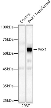
Western blot analysis of lysates from wild type (WT) and 293T cells transfected with PAX1 using PAX1 Rabbit pAb (CAB17513) at 1:400 dilution. Secondary antibody: HRP-conjugated Goat anti-Rabbit IgG (H+L) (CABS014) at 1:10000 dilution. Lysates/proteins: 25μg per lane. Blocking buffer: 3% nonfat dry milk in TBST. Detection: ECL Basic Kit (AbGn00020). Exposure time: 60s.