The PCCB Antibody (CAB5415) is a high-quality antibody developed for reliable detection and analysis of target proteins. This antibody, produced in rabbits, is highly specific to human samples and has been validated for use in Western blot applications. By targeting the PCCB protein, researchers can easily detect and analyze its expression in various cell types, making it an essential tool for studies in metabolic disorders, enzyme deficiencies, and other related research areas.The PCCB protein plays a critical role in maintaining metabolic homeostasis and energy production by catalyzing the final step in the breakdown of certain amino acids.
This antibody is validated for use in WB, ELISA applications and has demonstrated reactivity against Human, Mouse samples.
Product Name:
PCCB Antibody
SKU:
CAB5415
Size:
20μL, 100μL
Reactivity:
Human, Mouse
Conjugate:
Unconjugated
Immunogen:
Recombinant protein (or fragment).This information is considered to be commercially sensitive.
Recommended starting concentration is 1 μg/mL. Please optimize the concentration based on your specific assay requirements.
Synonyms:
PCCB
Positive Sample:
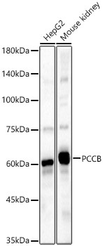
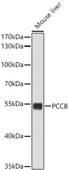
Hep G2, Mouse kidney, Mouse liver
Cellular Localization:
Mitochondrion Matrix.
Calculated MW:
58kDa
Observed MW:
54kDa/60kDa
The protein encoded by this gene is a subunit of the propionyl-CoA carboxylase (PCC) enzyme, which is involved in the catabolism of propionyl-CoA. PCC is a mitochondrial enzyme that probably acts as a dodecamer of six alpha subunits and six beta subunits. This gene encodes the beta subunit of PCC. Defects in this gene are a cause of propionic acidemia type II (PA-2). Multiple transcript variants encoding different isoforms have been found for this gene.
Purification Method
Affinity purification
Gene ID
5096
RRID
AB_2766223
Buffer Information
Store at -20℃. Avoid freeze / thaw cycles. Buffer: PBS containing 50% glycerol, preserved with proclin300 or sodium azide, pH 7.3.
Western blot analysis of various lysates, using PCCB Rabbit pAb (CAB5415) at 1:2000 dilution. Secondary antibody: HRP-conjugated Goat anti-Rabbit IgG (H+L) (CABS014) at 1:10000 dilution. Lysates/proteins: 25μg per lane. Blocking buffer: 3% nonfat dry milk in TBST. Detection: ECL Basic Kit (AbGn00020). Exposure time: 60s.
Western blot analysis of lysates from Mouse liver using PCCB Rabbit pAb (CAB5415) at 1:2000 dilution. Secondary antibody: HRP-conjugated Goat anti-Rabbit IgG (H+L) (CABS014) at 1:10000 dilution. Lysates/proteins: 25 μg per lane. Blocking buffer: 3% nonfat dry milk in TBST. Detection: ECL Basic Kit (AbGn00020). Exposure time: 30s.


")


