The PCDH1 Antibody (CAB10234) is a high-quality antibody developed for reliable detection and analysis of target proteins. This antibody, produced in rabbits, exhibits high reactivity towards human samples and has been validated for use in Western blot applications. By binding specifically to Protocadherin-1, this antibody allows for the detection and analysis of this protein in a variety of cell types, making it an excellent choice for studies in developmental biology and neuroscience.
This antibody is validated for use in WB, ELISA applications and has demonstrated reactivity against Human samples.
Product Name:
PCDH1 Antibody
SKU:
CAB10234
Size:
20μL, 100μL
Reactivity:
Human
Conjugate:
Unconjugated
Immunogen:
Recombinant protein (or fragment).This information is considered to be commercially sensitive.
Recommended starting concentration is 1 μg/mL. Please optimize the concentration based on your specific assay requirements.
Synonyms:
PC42, PCDH42, PCDH1
Positive Sample:
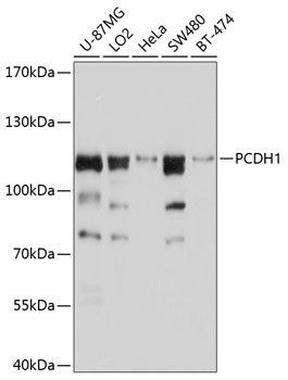
U-87MG, LO2, HeLa, SW480, BT-474
Cellular Localization:
Cell Junction, Cell Membrane, Single-Pass Type I Membrane Protein.
Calculated MW:
115kDa
Observed MW:
115kDa
This gene belongs to the protocadherin subfamily within the cadherin superfamily. The encoded protein is a membrane protein found at cell-cell boundaries. It is involved in neural cell adhesion, suggesting a possible role in neuronal development. The protein includes an extracelllular region, containing 7 cadherin-like domains, a transmembrane region and a C-terminal cytoplasmic region. Cells expressing the protein showed cell aggregation activity. Alternative splicing occurs in this gene.
Purification Method
Affinity purification
Gene ID
5097
RRID
AB_2757759
Buffer Information
Store at -20℃. Avoid freeze / thaw cycles. Buffer: PBS containing 50% glycerol, preserved with proclin300 or sodium azide, pH 7.3.
Western blot analysis of various lysates using PCDH1 Rabbit pAb (CAB10234) at 1:1000 dilution. Secondary antibody: HRP-conjugated Goat anti-Rabbit IgG (H+L) (CABS014) at 1:10000 dilution. Lysates/proteins: 25μg per lane. Blocking buffer: 3% nonfat dry milk in TBST. Detection: ECL Basic Kit (AbGn00020). Exposure time: 5s.