The PCGF6 Antibody (CAB5760) is a high-quality antibody developed for reliable detection and analysis of target proteins. This antibody, generated from rabbit hosts, exhibits high reactivity with human samples and is optimized for Western blot applications. By specifically binding to the PCGF6 protein, this antibody facilitates the detection and analysis of PCGF6 expression in various cell types, offering insight into its role in cellular processes.PCGF6, a component of the polycomb repressive complex 1 (PRC1), plays a crucial role in gene silencing and transcriptional regulation. Its involvement in chromatin dynamics and gene expression makes it a key player in cellular development and differentiation.
This antibody is validated for use in WB, ELISA applications and has demonstrated reactivity against Human, Mouse, Rat samples.
Product Name:
PCGF6 Antibody
SKU:
CAB5760
Size:
20μL, 100μL
Reactivity:
Human, Mouse, Rat
Conjugate:
Unconjugated
Immunogen:
Recombinant protein (or fragment).This information is considered to be commercially sensitive.
Recommended starting concentration is 1 μg/mL. Please optimize the concentration based on your specific assay requirements.
Synonyms:
MBLR, RNF134, PCGF6
Positive Sample:
MCF7
Cellular Localization:
Nucleus.
Calculated MW:
39kDa
Observed MW:
39kDa
The protein encoded by this gene contains a RING finger motif, which is most closely related to those of polycomb group (PcG) proteins RNF110/MEL-18 and BMI1. PcG proteins are known to form protein complexes and function as transcription repressors. This protein has been shown to interact with some PcG proteins and act as a transcription repressor. The activity of this protein is found to be regulated by cell cycle dependent phosphorylation. Alternatively spliced transcript variants encoding different isoforms have been identified.
Purification Method
Affinity purification
Gene ID
84108
RRID
AB_2766514
Buffer Information
Store at -20℃. Avoid freeze / thaw cycles. Buffer: PBS containing 50% glycerol, preserved with proclin300 or sodium azide, pH 7.3.
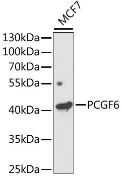
Western blot analysis of lysates from MCF7 cells, using PCGF6 Rabbit pAb (CAB5760) at 1:1000 dilution. Secondary antibody: HRP-conjugated Goat anti-Rabbit IgG (H+L) (CABS014) at 1:10000 dilution. Lysates/proteins: 25μg per lane. Blocking buffer: 3% nonfat dry milk in TBST. Detection: ECL Enhanced Kit (AbGn00021). Exposure time: 120s.