The PCK1 Antibody (CAB2036) is a high-quality antibody developed for reliable detection and analysis of target proteins. This enzyme plays a key role in gluconeogenesis, the process by which glucose is synthesized in the liver and kidneys. The antibody is raised in rabbits and is highly specific for human samples, making it ideal for Western blot applications.By targeting the PCK1 protein, researchers can investigate its role in regulating glucose production and metabolism, as well as its potential implications in conditions such as diabetes, obesity, and metabolic disorders.
This antibody is validated for use in WB, IF/ICC, IP, ELISA, IF-P applications and has demonstrated reactivity against Human, Mouse, Rat samples.
Product Name:
PCK1 Antibody
SKU:
CAB2036
Size:
20μL, 100μL
Reactivity:
Human, Mouse, Rat
Conjugate:
Unconjugated
Immunogen:
Recombinant protein (or fragment).This information is considered to be commercially sensitive.
0.5μg-4μg antibody for 400μg-600μg extracts of whole cells
IF/ICC
1:50 - 1:200
IF-P
1:50 - 1:200
ELISA
Recommended starting concentration is 1 μg/mL. Please optimize the concentration based on your specific assay requirements.
Synonyms:
PCKDC, PEPCK1, PEPCKC, PEPCK-C, PCK1
Positive Sample:
HepG2, Mouse liver, Mouse kidney, Rat liver, Rat pancreas, Rat kidney
Cellular Localization:
Cytoplasm.
Calculated MW:
69kDa
Observed MW:
69kDa
This gene is a main control point for the regulation of gluconeogenesis. The cytosolic enzyme encoded by this gene, along with GTP, catalyzes the formation of phosphoenolpyruvate from oxaloacetate, with the release of carbon dioxide and GDP. The expression of this gene can be regulated by insulin, glucocorticoids, glucagon, cAMP, and diet. Defects in this gene are a cause of cytosolic phosphoenolpyruvate carboxykinase deficiency. A mitochondrial isozyme of the encoded protein also has been characterized.
Purification Method
Affinity purification
Gene ID
5105
RRID
AB_2764060
Buffer Information
Store at -20℃. Avoid freeze / thaw cycles. Buffer: PBS containing 50% glycerol, preserved with proclin300 or sodium azide, pH 7.3.
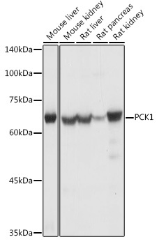
Western blot analysis of various lysates using PCK1 Rabbit pAb (CAB2036) at 1:1000 dilution. Secondary antibody: HRP-conjugated Goat anti-Rabbit IgG (H+L) (CABS014) at 1:10000 dilution. Lysates/proteins: 25μg per lane. Blocking buffer: 3% nonfat dry milk in TBST. Detection: ECL Basic Kit (AbGn00020). Exposure time: 1s.
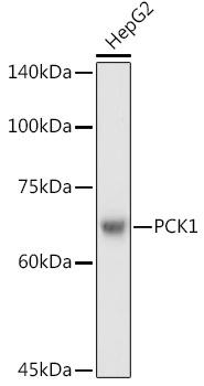
Western blot analysis of lysates from HepG2 cells, using PCK1 Rabbit pAb (CAB2036) at 1:1000 dilution. Secondary antibody: HRP-conjugated Goat anti-Rabbit IgG (H+L) (CABS014) at 1:10000 dilution. Lysates/proteins: 25μg per lane. Blocking buffer: 3% nonfat dry milk in TBST. Detection: ECL Basic Kit (AbGn00020). Exposure time: 3s.
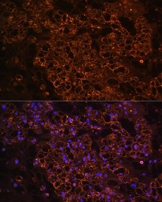
Immunofluorescence analysis of paraffin-embedded human kidney cancer using PCK1 Rabbit pAb (CAB2036) at dilution of 1:100 (40x lens). Secondary antibody: Cy3-conjugated Goat anti-Rabbit IgG (H+L) (CABS007) at 1:500 dilution. Blue: DAPI for nuclear staining.
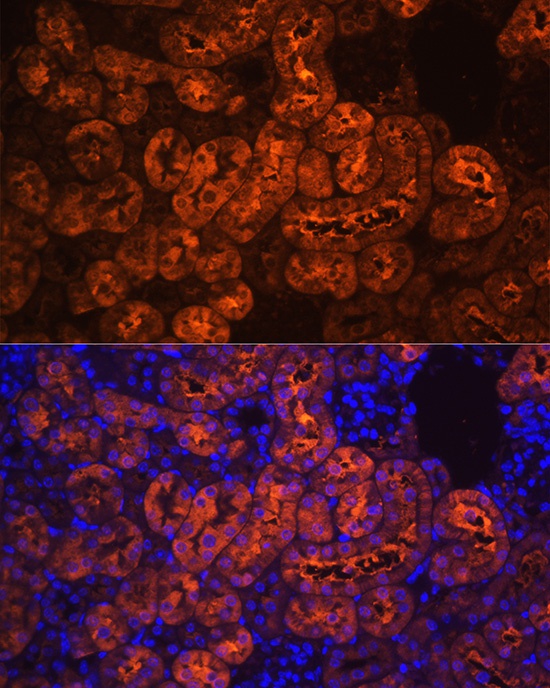
Immunofluorescence analysis of paraffin-embedded mouse kidney using PCK1 Rabbit pAb (CAB2036) at dilution of 1:100 (40x lens). Secondary antibody: Cy3-conjugated Goat anti-Rabbit IgG (H+L) (CABS007) at 1:500 dilution. Blue: DAPI for nuclear staining.
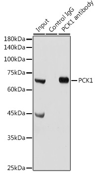
Immunoprecipitation analysis of 300 μg extracts of Mouse liver cells using 3 μg PCK1 antibody (CAB2036). Western blot was performed from the immunoprecipitate using PCK1 antibody (CAB2036) at a dilution of 1:1000.