The PCK1 Monoclonal Antibody (CAB22172) is a high-quality antibody developed for reliable detection and analysis of target proteins. This antibody, produced by Assay Genie, is specifically designed to target and identify PCK1 in various cell types and tissues, making it an ideal choice for studies in metabolic pathways, diabetes, and liver function.This monoclonal antibody, developed using hybridoma technology, is highly specific and sensitive, ensuring accurate and reliable results in experiments such as Western blotting and immunohistochemistry.
This antibody is validated for use in WB, ELISA applications and has demonstrated reactivity against Human, Mouse, Rat samples.
Product Name:
PCK1 Monoclonal Antibody
SKU:
CAB22172
Size:
20μL, 100μL
Reactivity:
Human, Mouse, Rat
Clone Number:
ARC56074
Conjugate:
Unconjugated
Immunogen:
Synthetic peptide. This information is considered to be commercially sensitive.
Recommended starting concentration is 1 μg/mL. Please optimize the concentration based on your specific assay requirements.
Synonyms:
PCKDC, PEPCK1, PEPCKC, PEPCK-C, PCK1
Positive Sample:
Hep G2, Mouse kidney, Rat kidney
Cellular Localization:
Cytoplasm.
Calculated MW:
69kDa
Observed MW:
69kDa
This gene is a main control point for the regulation of gluconeogenesis. The cytosolic enzyme encoded by this gene, along with GTP, catalyzes the formation of phosphoenolpyruvate from oxaloacetate, with the release of carbon dioxide and GDP. The expression of this gene can be regulated by insulin, glucocorticoids, glucagon, cAMP, and diet. Defects in this gene are a cause of cytosolic phosphoenolpyruvate carboxykinase deficiency. A mitochondrial isozyme of the encoded protein also has been characterized.
Purification Method
Affinity purification
Gene ID
5105
Buffer Information
Store at -20℃. Avoid freeze / thaw cycles. Buffer: PBS with 0.09% Sodium azide,0.05% BSA,50% glycerol,pH7.3.
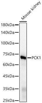
Western blot analysis of various lysates, using PCK1 Rabbit mAb (CAB22172) at1:2000 dilution. Secondary antibody: HRP-conjugated Goat anti-Rabbit IgG (H+L) (CABS014) at 1:10000 dilution. Lysates/proteins: 25μg per lane. Blocking buffer: 3% nonfat dry milk in TBST. Detection: ECL Basic Kit (AbGn00020). Exposure time: 0.5s.
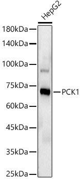
Western blot analysis of lysates from HepG2 cells, using PCK1 Rabbit mAb (CAB22172) at1:2000 dilution. Secondary antibody: HRP-conjugated Goat anti-Rabbit IgG (H+L) (CABS014) at 1:10000 dilution. Lysates/proteins: 25μg per lane. Blocking buffer: 3% nonfat dry milk in TBST. Detection: ECL Basic Kit (AbGn00020). Exposure time: 0.5s.
 at1:2000 dilution. Secondary antibody: HRP Goat Anti-Rabbit IgG (H+L) at 1:10000 dilution. Lysates/proteins: 25μg per lane. Blocking buffer: 3% nonfat dry milk in TBST.")
 at1:2000 dilution. Secondary antibody: HRP Goat Anti-Rabbit IgG (H+L) at 1:10000 dilution. Lysates/proteins: 25μg per lane. Blocking buffer: 3% nonfat dry milk in TBST.")

