The PDCD10 Antibody (CAB17094) is a high-quality antibody developed for reliable detection and analysis of target proteins. This gene encodes an evolutionarily conserved protein associated with cell apoptosis. The protein interacts with the serine/threonine protein kinase MST4 to modulate the extracellular signal-regulated kinase (ERK) pathway. It also interacts with and is phosphoryated by serine/threonine kinase 25, and is thought to function in a signaling pathway essential for vascular developent. Mutations in this gene are one cause of cerebral cavernous malformations, which are vascular malformations that cause seizures and cerebral hemorrhages. Multiple alternatively spliced variants, encoding the same protein, have been identified.
This antibody is validated for use in WB, IHC-P, IF/ICC, ELISA applications and has demonstrated reactivity against Human, Mouse, Rat samples.
Product Name:
PDCD10 Antibody
SKU:
CAB17094
Size:
100μL, 20μL
Reactivity:
Human, Mouse, Rat
Immunogen:
Recombinant protein (or fragment).This information is considered to be commercially sensitive.
Tested Applications:
WBIHC-PIF/ICCELISA
Recommended Dilution:
WB
1:500 - 1:1000
IHC-P
1:50 - 1:200
IF/ICC
1:50 - 1:200
ELISA
Recommended starting concentration is 1 μg/mL. Please optimize the concentration based on your specific assay requirements.
This gene encodes an evolutionarily conserved protein associated with cell apoptosis. The protein interacts with the serine/threonine protein kinase MST4 to modulate the extracellular signal-regulated kinase (ERK) pathway. It also interacts with and is phosphoryated by serine/threonine kinase 25, and is thought to function in a signaling pathway essential for vascular developent. Mutations in this gene are one cause of cerebral cavernous malformations, which are vascular malformations that cause seizures and cerebral hemorrhages. Multiple alternatively spliced variants, encoding the same protein, have been identified.
Purification Method
Affinity purification
Gene ID
11235
RRID
AB_2770821
Buffer Information
Store at -20℃. Avoid freeze / thaw cycles. Buffer: PBS containing 50% glycerol, preserved with proclin300 or sodium azide, pH 7.3.
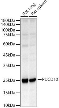
Western blot analysis of various lysates, using PDCD10 Rabbit pAb (CAB17094) at 1:500 dilution. Secondary antibody: HRP-conjugated Goat anti-Rabbit IgG (H+L) (AS014) at 1:10000 dilution. Lysates/proteins: 25μg per lane. Blocking buffer: 3% nonfat dry milk in TBST. Detection: ECL Basic Kit (AbGn00020). Exposure time: 45s.
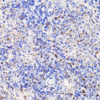
Immunohistochemistry analysis of paraffin-embedded Rat spleen using PDCD10 Rabbit pAb (CAB17094) at dilution of 1:100 (40x lens). Microwave antigen retrieval performed with 0.01M PBS Buffer (pH 7.2) prior to IHC staining.
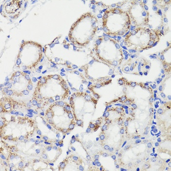
Immunohistochemistry analysis of paraffin-embedded Mouse kidney using PDCD10 Rabbit pAb (CAB17094) at dilution of 1:100 (40x lens). Microwave antigen retrieval performed with 0.01M PBS Buffer (pH 7.2) prior to IHC staining.
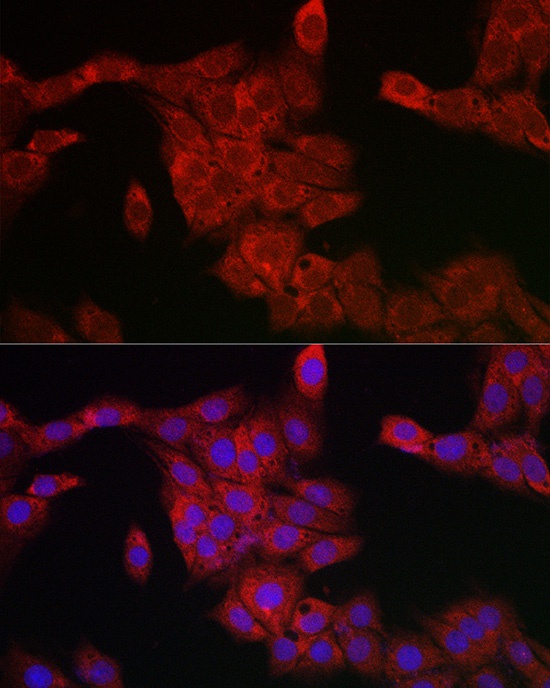
Immunofluorescence analysis of PC-12 cells using PDCD10 Rabbit pAb (CAB17094) at dilution of 1:200 (40x lens). Secondary antibody: Cy3-conjugated Goat anti-Rabbit IgG (H+L) (AS007) at 1:500 dilution. Blue: DAPI for nuclear staining.