The PDE12 Antibody (CAB17270) is a high-quality antibody developed for reliable detection and analysis of target proteins. This rabbit polyclonal antibody is highly specific for human samples and has been validated for use in Western blotting applications. By binding to the PDE12 enzyme, this antibody allows for the detection and analysis of PDE12 expression in various cell types, making it a valuable tool for studies in molecular biology and drug development.
This antibody is validated for use in WB, IF/ICC, ELISA applications and has demonstrated reactivity against Mouse, Rat samples.
Product Name:
PDE12 Antibody
SKU:
CAB17270
Size:
20μL, 100μL
Reactivity:
Mouse, Rat
Immunogen:
Recombinant protein (or fragment).This information is considered to be commercially sensitive.
Recommended starting concentration is 1 μg/mL. Please optimize the concentration based on your specific assay requirements.
Synonyms:
2-PDE, 2'-PDE, PDE12
Positive Sample:
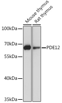
Mouse thymus, Rat thymus
Cellular Localization:
Cytosol, Mitochondrial Matrix, Mitochondrion.
Calculated MW:
67kDa
Observed MW:
67kDa
Enables 3'-5'-exoribonuclease activity. Involved in several processes, including RNA metabolic process; cellular response to cytokine stimulus; and regulation of mitochondrial mRNA stability. Located in mitochondrial matrix.
Purification Method
Affinity purification
Gene ID
201626
RRID
AB_2770823
Buffer Information
Store at -20℃. Avoid freeze / thaw cycles. Buffer: PBS with 0.01% thimerosal,50% glycerol,pH7.3.
Western blot analysis of various lysates using PDE12 Rabbit pAb (CAB17270) at 1:1000 dilution. Secondary antibody: HRP-conjugated Goat anti-Rabbit IgG (H+L) (CABS014) at 1:10000 dilution. Lysates/proteins: 25μg per lane. Blocking buffer: 3% nonfat dry milk in TBST. Detection: ECL Basic Kit (AbGn00020). Exposure time: 10s.
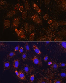
Immunofluorescence analysis of C6 cells using PDE12 Rabbit pAb (CAB17270) at dilution of 1:100. Secondary antibody: Cy3-conjugated Goat anti-Rabbit IgG (H+L) (CABS007) at 1:500 dilution. Blue: DAPI for nuclear staining.