The PDE3B Antibody (CAB8448) is a high-quality antibody developed for reliable detection and analysis of target proteins. This antibody, raised in rabbits, is highly specific to human PDE3B and is validated for use in Western blot applications. By binding to the PDE3B protein, this antibody enables precise detection and analysis in a variety of cell types, making it an essential tool for investigations in the fields of metabolism, cardiovascular biology, and cancer research.PDE3B plays a crucial role in cellular signaling pathways that regulate metabolism, cardiac function, and smooth muscle contraction.
This antibody is validated for use in WB, ELISA applications and has demonstrated reactivity against Human, Mouse samples.
Product Name:
PDE3B Antibody
SKU:
CAB8448
Size:
20μL, 100μL
Reactivity:
Human, Mouse
Conjugate:
Unconjugated
Immunogen:
Recombinant protein (or fragment).This information is considered to be commercially sensitive.
Recommended starting concentration is 1 μg/mL. Please optimize the concentration based on your specific assay requirements.
Synonyms:
HcGIP1, cGIPDE1, PDE3B
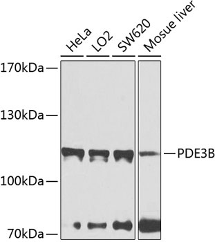
Positive Sample:
HeLa, LO2, SW620, Mosue liver
Cellular Localization:
Membrane, Multi-Pass Membrane Protein.
Calculated MW:
124kDa
Observed MW:
115kDa
Enables 3',5'-cyclic-nucleotide phosphodiesterase activity. Involved in negative regulation of angiogenesis; negative regulation of cell adhesion; and negative regulation of lipid catabolic process. Located in membrane. Part of guanyl-nucleotide exchange factor complex.
Purification Method
Affinity purification
Gene ID
5140
RRID
AB_2770825
Buffer Information
Store at -20℃. Avoid freeze / thaw cycles. Buffer: PBS containing 50% glycerol, preserved with proclin300 or sodium azide, pH 7.3.
Western blot analysis of various lysates using PDE3B Rabbit pAb (CAB8448) at 1:1000 dilution. Secondary antibody: HRP-conjugated Goat anti-Rabbit IgG (H+L) (CABS014) at 1:10000 dilution. Lysates/proteins: 25μg per lane. Blocking buffer: 3% nonfat dry milk in TBST. Detection: ECL Basic Kit (AbGn00020). Exposure time: 40s.