The PDE7A Antibody (CAB15079) is a high-quality antibody developed for reliable detection and analysis of target proteins. This antibody, produced in rabbits, specifically targets the PDE7A protein and is highly reactive with human samples. Validated for use in Western blot applications, the PDE7A Polyclonal Antibody allows for the detection and analysis of PDE7A in various cell types.PDE7A is known to play a role in a variety of physiological processes, including immune response, inflammation, and neuronal signaling. Dysregulation of PDE7A has been linked to various diseases, making it an attractive target for drug discovery and therapeutic intervention.
This antibody is validated for use in WB, ELISA applications and has demonstrated reactivity against Human, Mouse, Rat samples.
Product Name:
PDE7A Antibody
SKU:
CAB15079
Size:
20μL, 100μL
Reactivity:
Human, Mouse, Rat
Conjugate:
Unconjugated
Immunogen:
Recombinant protein (or fragment).This information is considered to be commercially sensitive.
Recommended starting concentration is 1 μg/mL. Please optimize the concentration based on your specific assay requirements.
Synonyms:
HCP1, PDE7, PDE7A
Positive Sample:
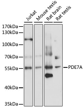
Jurkat, mouse testis, rat brain, rat testis
Cellular Localization:
Cytoplasm, Cytosol.
Calculated MW:
56kDa
Observed MW:
56kDa
The protein encoded by this gene belongs to the cyclic nucleotide phosphodiesterase (PDE) family, and PDE7 subfamily. This PDE hydrolyzes the second messenger, cAMP, which is a regulator and mediator of a number of cellular responses to extracellular signals. Thus, by regulating the cellular concentration of cAMP, this protein plays a key role in many important physiological processes. Alternatively spliced transcript variants encoding different isoforms have been described for this gene.
Purification Method
Affinity purification
Gene ID
5150
RRID
AB_2761961
Buffer Information
Store at -20℃. Avoid freeze / thaw cycles. Buffer: PBS with 0.01% thimerosal,50% glycerol,pH7.3.
Western blot analysis of various lysates using PDE7A Rabbit pAb (CAB15079) at 1:1000 dilution. Secondary antibody: HRP-conjugated Goat anti-Rabbit IgG (H+L) (CABS014) at 1:10000 dilution. Lysates/proteins: 25μg per lane. Blocking buffer: 3% nonfat dry milk in TBST. Detection: ECL Enhanced Kit (AbGn00021). Exposure time: 10s.