The PDGFRB Monoclonal Antibody (CAB19531) is a high-quality antibody developed for reliable detection and analysis of target proteins. This antibody, generated in rabbits, is highly specific to human samples and has been rigorously validated for use in Western blot applications.The PDGF receptor beta is a vital component in several cellular processes, including wound healing, tissue repair, and angiogenesis. Dysregulation of this receptor has been implicated in various diseases, making it a prime target for therapeutic interventions.
This antibody is validated for use in WB, IF/ICC, ELISA applications and has demonstrated reactivity against Human, Mouse samples.
Product Name:
PDGFRB Monoclonal Antibody
SKU:
CAB19531
Size:
20μL, 100μL
Reactivity:
Human, Mouse
Clone Number:
ARC0009
Conjugate:
Unconjugated
Immunogen:
Synthetic peptide. This information is considered to be commercially sensitive.
Cell Membrane, Cytoplasmic Vesicle, Lysosome Lumen, Single-Pass Type I Membrane Protein.
Calculated MW:
124kDa
Observed MW:
190kDa
The protein encoded by this gene is a cell surface tyrosine kinase receptor for members of the platelet-derived growth factor family. These growth factors are mitogens for cells of mesenchymal origin. The identity of the growth factor bound to a receptor monomer determines whether the functional receptor is a homodimer (PDGFB or PDGFD) or a heterodimer (PDGFA and PDGFB). This gene is essential for normal development of the cardiovascular system and aids in rearrangement of the actin cytoskeleton. This gene is flanked on chromosome 5 by the genes for granulocyte-macrophage colony-stimulating factor and macrophage-colony stimulating factor receptor; all three genes may be implicated in the 5-q syndrome. A translocation between chromosomes 5 and 12, that fuses this gene to that of the ETV6 gene, results in chronic myeloproliferative disorder with eosinophilia.
Purification Method
Affinity purification
Gene ID
5159
RRID
AB_2832987
Buffer Information
Store at -20℃. Avoid freeze / thaw cycles. Buffer: PBS containing 50% glycerol and 0.05% BSA, preserved with proclin300 or sodium azide, pH 7.3.
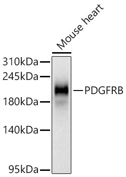
Western blot analysis of lysates from Mouse heart using PDGFRB Rabbit mAb (CAB19531) at 1:1000 dilution incubated overnight at 4℃. Secondary antibody: HRP-conjugated Goat anti-Rabbit IgG (H+L) (CABS014) at 1:10000 dilution. Lysates/proteins: 25 μg per lane. Blocking buffer: 3% nonfat dry milk in TBST. Detection: ECL Basic Kit (AbGn00020). Exposure time: 30s.
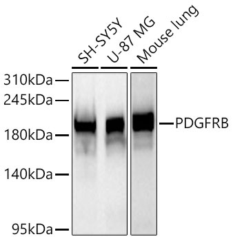
Western blot analysis of various lysates using PDGFRB Rabbit mAb (CAB19531) at 1:1000 dilution incubated overnight at 4℃. Secondary antibody: HRP-conjugated Goat anti-Rabbit IgG (H+L) (CABS014) at 1:10000 dilution. Lysates/proteins: 25 μg per lane. Blocking buffer: 3% nonfat dry milk in TBST. Detection: ECL Basic Kit (AbGn00020). Exposure time: 20s.